Alessandro Moreira Freire, Orlando Aguirre Guedes, Lara Borges de Deus, Paulo Eduardo Mafra, Gustavo Silva Chaves, Karolina Kellen Matias, Juliano Gonçalves Miguel, Erica Eugênia Javarez Freire, Daniel de Almeida Decurcio
{"title":"Endoaesthetic Management of Type II Dens Invaginatus Associated With Root Perforation and Apical Abscess: A Case Report.","authors":"Alessandro Moreira Freire, Orlando Aguirre Guedes, Lara Borges de Deus, Paulo Eduardo Mafra, Gustavo Silva Chaves, Karolina Kellen Matias, Juliano Gonçalves Miguel, Erica Eugênia Javarez Freire, Daniel de Almeida Decurcio","doi":"10.1155/crid/2909252","DOIUrl":null,"url":null,"abstract":"<p><p>Dens invaginatus (DI) is a developmental anomaly that affects teeth. This case report demonstrates the successful endoaesthetic management of a Type II DI in the maxillary right lateral incisor (Tooth 12). A 12-year-old female patient presented with pain and swelling in Tooth 12, which had previously been diagnosed with DI and accessed endodontically. The patient was also concerned about the aesthetic appearance of her anterior teeth. Clinical, radiographic, and tomographic findings confirmed Oehler's type II DI, with root perforation and an apical abscess in Tooth 12, as well as pulp necrosis in Tooth 13. Nonsurgical root canal treatment was recommended for both teeth, with additional root perforation repair for Tooth 12. Multiple visits were required for nonsurgical endodontic therapy. During the initial visit, the perforation was identified and sealed, followed by chemomechanical preparation and placement of calcium hydroxide paste. Ten months later, the root canals were filled. After endodontic treatment, the patient underwent in-office bleaching, followed by composite resin veneers on the upper anterior teeth, using the preformed metal matrix technique. This resulted in excellent aesthetic outcomes. At the 2-year follow-up examination, the patient remained asymptomatic, and radiographic assessment showed complete resolution of the periapical lesion. This case highlights the importance of multidisciplinary collaboration, precise treatment planning, and patient-centered care in achieving optimal endoaesthetic results in complex cases.</p>","PeriodicalId":46841,"journal":{"name":"Case Reports in Dentistry","volume":"2025 ","pages":"2909252"},"PeriodicalIF":0.9000,"publicationDate":"2025-04-28","publicationTypes":"Journal Article","fieldsOfStudy":null,"isOpenAccess":false,"openAccessPdf":"https://www.ncbi.nlm.nih.gov/pmc/articles/PMC12052460/pdf/","citationCount":"0","resultStr":null,"platform":"Semanticscholar","paperid":null,"PeriodicalName":"Case Reports in Dentistry","FirstCategoryId":"1085","ListUrlMain":"https://doi.org/10.1155/crid/2909252","RegionNum":0,"RegionCategory":null,"ArticlePicture":[],"TitleCN":null,"AbstractTextCN":null,"PMCID":null,"EPubDate":"2025/1/1 0:00:00","PubModel":"eCollection","JCR":"Q4","JCRName":"DENTISTRY, ORAL SURGERY & MEDICINE","Score":null,"Total":0}
引用次数: 0
Abstract
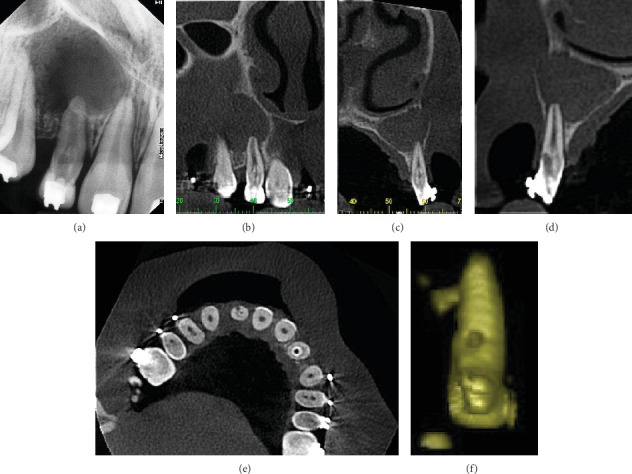
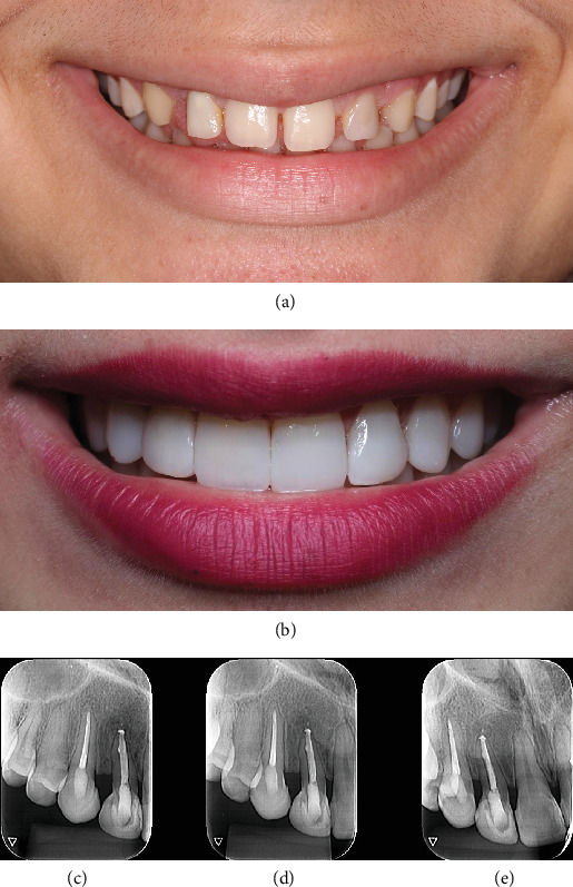
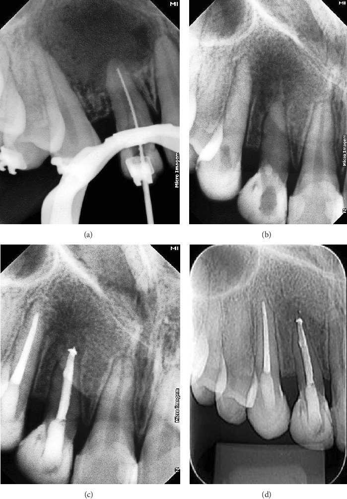
Dens invaginatus (DI) is a developmental anomaly that affects teeth. This case report demonstrates the successful endoaesthetic management of a Type II DI in the maxillary right lateral incisor (Tooth 12). A 12-year-old female patient presented with pain and swelling in Tooth 12, which had previously been diagnosed with DI and accessed endodontically. The patient was also concerned about the aesthetic appearance of her anterior teeth. Clinical, radiographic, and tomographic findings confirmed Oehler's type II DI, with root perforation and an apical abscess in Tooth 12, as well as pulp necrosis in Tooth 13. Nonsurgical root canal treatment was recommended for both teeth, with additional root perforation repair for Tooth 12. Multiple visits were required for nonsurgical endodontic therapy. During the initial visit, the perforation was identified and sealed, followed by chemomechanical preparation and placement of calcium hydroxide paste. Ten months later, the root canals were filled. After endodontic treatment, the patient underwent in-office bleaching, followed by composite resin veneers on the upper anterior teeth, using the preformed metal matrix technique. This resulted in excellent aesthetic outcomes. At the 2-year follow-up examination, the patient remained asymptomatic, and radiographic assessment showed complete resolution of the periapical lesion. This case highlights the importance of multidisciplinary collaboration, precise treatment planning, and patient-centered care in achieving optimal endoaesthetic results in complex cases.
期刊介绍:
Case Reports in Dentistry is a peer-reviewed, Open Access journal that publishes case reports and case series in all areas of dentistry, including periodontal diseases, dental implants, oral pathology, as well as oral and maxillofacial surgery.

 求助内容:
求助内容: 应助结果提醒方式:


