Mila Ćetković, Jelena Boljanović, Ema Bexheti, Filip Vitošević, Damljan Bogićević, Sonja Milašinović, Sadi Bexheti, Dejan Ćetković, Aleksandra Dožić, Milan Milisavljević
{"title":"Fenestrations of cerebral arteries and their correlation with brain aneurysms.","authors":"Mila Ćetković, Jelena Boljanović, Ema Bexheti, Filip Vitošević, Damljan Bogićević, Sonja Milašinović, Sadi Bexheti, Dejan Ćetković, Aleksandra Dožić, Milan Milisavljević","doi":"10.3389/fnana.2025.1523305","DOIUrl":null,"url":null,"abstract":"<p><p>Fenestration of the intracranial artery is an anatomical remnant from the embryonic development of the vascular system. A cerebral aneurysm is a focal pathological dilation of the arterial wall. The occurrence of an aneurysm at the site of fenestration is rare in cerebral circulation but may have potential clinical implications. This study aimed to identify the frequencies of fenestrations and aneurysms, their locations, and their relationships. The vasculature of 35 adult brains was used for micromorphological dissection and analysis under a stereoscopic microscope, following an arterial injection with a mixture of formaldehyde, melted gelatin, and the solution of India ink. Additionally, we analyzed another group of vascular casts obtained from 15 brains injected with methyl methacrylate (MMA). A fenestration of the M1 segment of the middle cerebral artery (MCA) was sectioned for histological analysis. We also examined computed tomography (CT) angiograms of 1,230 patients, analyzed the data, and compared the findings with anatomical observations. In our group of 50 anatomical specimens, fenestrations were found in 12 brains (24%), affecting different cerebral arteries, with three cases showing double fenestrations on the same vessel. Aneurysms were observed in six brains (12%), always one per brain, with one case (2.00%) involving an aneurysm associated with the wall of a fenestration. Analysis of CT angiograms from 1,230 patients showed 26 arterial fenestrations (2.11%) in 26 patients, 28 aneurysms (2.28%), and one case (0.08%) where an aneurysm arose from a fenestration. The presence of an aneurysm on a fenestrated cerebral artery is a rare phenomenon, occurring far less frequently than isolated fenestrations or aneurysm formation.</p>","PeriodicalId":12572,"journal":{"name":"Frontiers in Neuroanatomy","volume":"19 ","pages":"1523305"},"PeriodicalIF":2.3000,"publicationDate":"2025-03-26","publicationTypes":"Journal Article","fieldsOfStudy":null,"isOpenAccess":false,"openAccessPdf":"https://www.ncbi.nlm.nih.gov/pmc/articles/PMC11979215/pdf/","citationCount":"0","resultStr":null,"platform":"Semanticscholar","paperid":null,"PeriodicalName":"Frontiers in Neuroanatomy","FirstCategoryId":"3","ListUrlMain":"https://doi.org/10.3389/fnana.2025.1523305","RegionNum":4,"RegionCategory":"医学","ArticlePicture":[],"TitleCN":null,"AbstractTextCN":null,"PMCID":null,"EPubDate":"2025/1/1 0:00:00","PubModel":"eCollection","JCR":"Q1","JCRName":"ANATOMY & MORPHOLOGY","Score":null,"Total":0}
引用次数: 0
Abstract
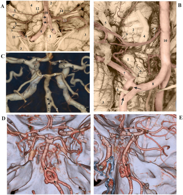
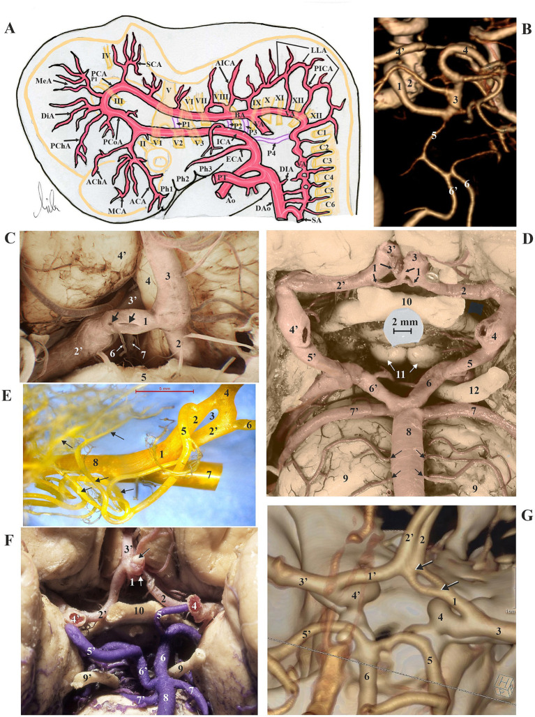
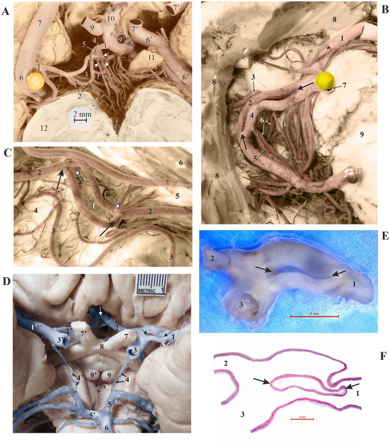
Fenestration of the intracranial artery is an anatomical remnant from the embryonic development of the vascular system. A cerebral aneurysm is a focal pathological dilation of the arterial wall. The occurrence of an aneurysm at the site of fenestration is rare in cerebral circulation but may have potential clinical implications. This study aimed to identify the frequencies of fenestrations and aneurysms, their locations, and their relationships. The vasculature of 35 adult brains was used for micromorphological dissection and analysis under a stereoscopic microscope, following an arterial injection with a mixture of formaldehyde, melted gelatin, and the solution of India ink. Additionally, we analyzed another group of vascular casts obtained from 15 brains injected with methyl methacrylate (MMA). A fenestration of the M1 segment of the middle cerebral artery (MCA) was sectioned for histological analysis. We also examined computed tomography (CT) angiograms of 1,230 patients, analyzed the data, and compared the findings with anatomical observations. In our group of 50 anatomical specimens, fenestrations were found in 12 brains (24%), affecting different cerebral arteries, with three cases showing double fenestrations on the same vessel. Aneurysms were observed in six brains (12%), always one per brain, with one case (2.00%) involving an aneurysm associated with the wall of a fenestration. Analysis of CT angiograms from 1,230 patients showed 26 arterial fenestrations (2.11%) in 26 patients, 28 aneurysms (2.28%), and one case (0.08%) where an aneurysm arose from a fenestration. The presence of an aneurysm on a fenestrated cerebral artery is a rare phenomenon, occurring far less frequently than isolated fenestrations or aneurysm formation.
期刊介绍:
Frontiers in Neuroanatomy publishes rigorously peer-reviewed research revealing important aspects of the anatomical organization of all nervous systems across all species. Specialty Chief Editor Javier DeFelipe at the Cajal Institute (CSIC) is supported by an outstanding Editorial Board of international experts. This multidisciplinary open-access journal is at the forefront of disseminating and communicating scientific knowledge and impactful discoveries to researchers, academics, clinicians and the public worldwide.

 求助内容:
求助内容: 应助结果提醒方式:


