Woohyun Go, Wonkyu Park, Gunha Hwang, Soyon An, Hee Chun Lee, Tae Sung Hwang
{"title":"Cor triatriatum sinister with partial atrioventricular septal defect in a cat.","authors":"Woohyun Go, Wonkyu Park, Gunha Hwang, Soyon An, Hee Chun Lee, Tae Sung Hwang","doi":"10.17221/91/2024-VETMED","DOIUrl":null,"url":null,"abstract":"<p><p>A 6-year-old female neutered Turkish Angora cat was referred due to tachypnoea. The patient was diagnosed with cardiomegaly at a local hospital during a health screening two years ago. Tachypnoea occurred one year ago. On physical examination, the patient presented with a respiratory rate of 72 breaths per minute and a systolic blood pressure of 70 mmHg. Thoracic radiographs revealed severe cardiomegaly, left atrium (LA) enlargement, right atrium (RA) enlargement, right ventricular enlargement, and dilation of pulmonary arteries and veins. An alveolar pattern was identified in the right and left cranial lung lobes. Echocardiography revealed a membrane that divided the LA into two chambers, a defect in the lower atrial septum, and elongation of anterior or posterior tricuspid valves (TV). However, septal TV was not observed. During systole, blood flow from LA to RA was confirmed through a defect in the atrial septum. During diastole, blood flow from LA to left ventricular was confirmed. These findings suggest cor triatriatum sinister (CTS) with partial atrioventricular septal defect (AVSD). This report describes echocardiographic diagnosis of CTS with partial AVSD in a cat.</p>","PeriodicalId":23532,"journal":{"name":"Veterinarni Medicina","volume":"70 3","pages":"110-115"},"PeriodicalIF":0.8000,"publicationDate":"2025-03-24","publicationTypes":"Journal Article","fieldsOfStudy":null,"isOpenAccess":false,"openAccessPdf":"https://www.ncbi.nlm.nih.gov/pmc/articles/PMC12001872/pdf/","citationCount":"0","resultStr":null,"platform":"Semanticscholar","paperid":null,"PeriodicalName":"Veterinarni Medicina","FirstCategoryId":"97","ListUrlMain":"https://doi.org/10.17221/91/2024-VETMED","RegionNum":4,"RegionCategory":"农林科学","ArticlePicture":[],"TitleCN":null,"AbstractTextCN":null,"PMCID":null,"EPubDate":"2025/3/1 0:00:00","PubModel":"eCollection","JCR":"Q3","JCRName":"VETERINARY SCIENCES","Score":null,"Total":0}
引用次数: 0
Abstract
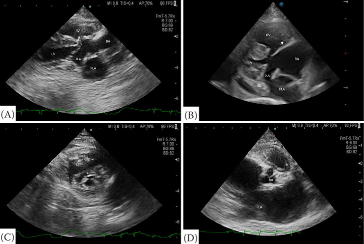
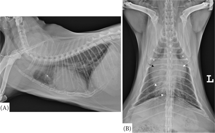
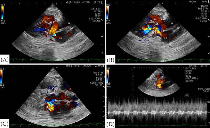
A 6-year-old female neutered Turkish Angora cat was referred due to tachypnoea. The patient was diagnosed with cardiomegaly at a local hospital during a health screening two years ago. Tachypnoea occurred one year ago. On physical examination, the patient presented with a respiratory rate of 72 breaths per minute and a systolic blood pressure of 70 mmHg. Thoracic radiographs revealed severe cardiomegaly, left atrium (LA) enlargement, right atrium (RA) enlargement, right ventricular enlargement, and dilation of pulmonary arteries and veins. An alveolar pattern was identified in the right and left cranial lung lobes. Echocardiography revealed a membrane that divided the LA into two chambers, a defect in the lower atrial septum, and elongation of anterior or posterior tricuspid valves (TV). However, septal TV was not observed. During systole, blood flow from LA to RA was confirmed through a defect in the atrial septum. During diastole, blood flow from LA to left ventricular was confirmed. These findings suggest cor triatriatum sinister (CTS) with partial atrioventricular septal defect (AVSD). This report describes echocardiographic diagnosis of CTS with partial AVSD in a cat.
期刊介绍:
The journal Veterinarni Medicina publishes in English original papers, short communications, critical reviews and case reports from all fields of veterinary and biomedical sciences.
 求助内容:
求助内容: 应助结果提醒方式:


