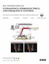
M Nicole MacMullin, Theresa Gu, Thomas G Landry, Nicholas Campbell, Sean D Christie, Jeremy A Brown
{"title":"A High Frequency Ultrasound Endoscope for Minimally Invasive Spine Surgery.","authors":"M Nicole MacMullin, Theresa Gu, Thomas G Landry, Nicholas Campbell, Sean D Christie, Jeremy A Brown","doi":"10.1109/TUFFC.2025.3559870","DOIUrl":null,"url":null,"abstract":"<p><p>The transition to minimally invasive spinal surgery over traditional open procedures requires the development of imaging techniques that meet the size constraints. Due to size restrictions associated with minimally invasive spine surgery, current imaging techniques are largely limited to microscopy, which is only capable of line-of-sight imaging of the tissue surface. A miniature, high-resolution ultrasound imaging endoscope has been developed as a potential alternative imaging method that would enable intra-operative guidance. We have designed and developed a 30 MHz miniature, 64-element, high-resolution imaging endoscope using PIN-PMT-PT single crystal as the piezoelectric substrate. The packaged probe had cross-sectional dimensions of 3.8 mm x 4.2 mm and a length of 14 cm. Two editions of the endoscope were created with a forward-facing and 40⁰ angle to enable visualization of structures during a medial and lateral approach, respectively. The probe was combined with a custom imaging system that produced real-time images with a field of view ranging between ±32⁰ and an image depth of 15 mm. The two-way axial resolution was measured to be 38 μm based on the -6 dB width of the pulse envelope. The -6 dB lateral resolution was measured to be 113 μm, 131 μm, and 158 μm at steering angles of 0⁰, 12⁰, and 25⁰ respectively, which were close to the simulated values of 106 μm, 118 μm and 144 μm. Preliminary clinical imaging studies successfully demonstrated the visualization of pertinent spinal anatomy during minimally invasive surgeries. The imaging probe was also able to demonstrate compression and decompression of nerve roots, supporting its potential use as a clinical tool.</p>","PeriodicalId":13322,"journal":{"name":"IEEE transactions on ultrasonics, ferroelectrics, and frequency control","volume":"PP ","pages":""},"PeriodicalIF":3.0000,"publicationDate":"2025-04-10","publicationTypes":"Journal Article","fieldsOfStudy":null,"isOpenAccess":false,"openAccessPdf":"","citationCount":"0","resultStr":null,"platform":"Semanticscholar","paperid":null,"PeriodicalName":"IEEE transactions on ultrasonics, ferroelectrics, and frequency control","FirstCategoryId":"5","ListUrlMain":"https://doi.org/10.1109/TUFFC.2025.3559870","RegionNum":2,"RegionCategory":"工程技术","ArticlePicture":[],"TitleCN":null,"AbstractTextCN":null,"PMCID":null,"EPubDate":"","PubModel":"","JCR":"Q1","JCRName":"ACOUSTICS","Score":null,"Total":0}
引用次数: 0
Abstract
The transition to minimally invasive spinal surgery over traditional open procedures requires the development of imaging techniques that meet the size constraints. Due to size restrictions associated with minimally invasive spine surgery, current imaging techniques are largely limited to microscopy, which is only capable of line-of-sight imaging of the tissue surface. A miniature, high-resolution ultrasound imaging endoscope has been developed as a potential alternative imaging method that would enable intra-operative guidance. We have designed and developed a 30 MHz miniature, 64-element, high-resolution imaging endoscope using PIN-PMT-PT single crystal as the piezoelectric substrate. The packaged probe had cross-sectional dimensions of 3.8 mm x 4.2 mm and a length of 14 cm. Two editions of the endoscope were created with a forward-facing and 40⁰ angle to enable visualization of structures during a medial and lateral approach, respectively. The probe was combined with a custom imaging system that produced real-time images with a field of view ranging between ±32⁰ and an image depth of 15 mm. The two-way axial resolution was measured to be 38 μm based on the -6 dB width of the pulse envelope. The -6 dB lateral resolution was measured to be 113 μm, 131 μm, and 158 μm at steering angles of 0⁰, 12⁰, and 25⁰ respectively, which were close to the simulated values of 106 μm, 118 μm and 144 μm. Preliminary clinical imaging studies successfully demonstrated the visualization of pertinent spinal anatomy during minimally invasive surgeries. The imaging probe was also able to demonstrate compression and decompression of nerve roots, supporting its potential use as a clinical tool.
期刊介绍:
IEEE Transactions on Ultrasonics, Ferroelectrics and Frequency Control includes the theory, technology, materials, and applications relating to: (1) the generation, transmission, and detection of ultrasonic waves and related phenomena; (2) medical ultrasound, including hyperthermia, bioeffects, tissue characterization and imaging; (3) ferroelectric, piezoelectric, and piezomagnetic materials, including crystals, polycrystalline solids, films, polymers, and composites; (4) frequency control, timing and time distribution, including crystal oscillators and other means of classical frequency control, and atomic, molecular and laser frequency control standards. Areas of interest range from fundamental studies to the design and/or applications of devices and systems.