Abdullah S Alqahtani, Wasan M Alshareef, Hanan T Aljadani, Wesal O Hawsawi, Marya H Shaheen
{"title":"The efficacy of artificial intelligence in diabetic retinopathy screening: a systematic review and meta-analysis.","authors":"Abdullah S Alqahtani, Wasan M Alshareef, Hanan T Aljadani, Wesal O Hawsawi, Marya H Shaheen","doi":"10.1186/s40942-025-00670-9","DOIUrl":null,"url":null,"abstract":"<p><strong>Background: </strong>To evaluate the efficacy of artificial intelligence (AI) in screening for diabetic retinopathy (DR) using fundus images and optical coherence tomography (OCT) in comparison to traditional screening methods.</p><p><strong>Methods: </strong>This systematic review was registered with PROSPERO (ID: CRD42024560750). Systematic searches were conducted in PubMed Medline, Cochrane Central, ScienceDirect, and Web of Science using keywords such as \"diabetic retinopathy,\" \"screening,\" and \"artificial intelligence.\" Only studies published in English from 2019 to July 22, 2024, were considered. We also manually reviewed the reference lists of relevant reviews. Two independent reviewers assessed the risk of bias using the QUADAS-2 tool, resolving disagreements through discussion with the principal investigator. Meta-analysis was performed using MetaDiSc software (version 1.4). To calculate combined sensitivity, specificity, summary receiver operating characteristic (SROC) plots, forest plots, and subgroup analyses were performed according to clinician type (ophthalmologists vs. retina specialists) and imaging modality (fundus images vs. fundus images + OCT).</p><p><strong>Results: </strong>18 studies were included. Meta-analysis showed that AI systems demonstrated superior diagnostic performance compared to doctors, with the pooled sensitivity, specificity, diagnostic odds ratio, and Cochrane Q index of the AI being 0.877, 0.906, 0.94, and 153.79 accordingly. The Fagan nomogram analysis further confirmed the strong diagnostic value of AI. Subgroup analyses revealed that factors like imaging modality, and doctor expertise can influence diagnostic performance.</p><p><strong>Conclusion: </strong>AI systems have demonstrated strong diagnostic performance in detecting diabetic retinopathy, with sensitivity and specificity comparable to or exceeding traditional clinicians.</p>","PeriodicalId":14289,"journal":{"name":"International Journal of Retina and Vitreous","volume":"11 1","pages":"48"},"PeriodicalIF":2.4000,"publicationDate":"2025-04-22","publicationTypes":"Journal Article","fieldsOfStudy":null,"isOpenAccess":false,"openAccessPdf":"https://www.ncbi.nlm.nih.gov/pmc/articles/PMC12012971/pdf/","citationCount":"0","resultStr":null,"platform":"Semanticscholar","paperid":null,"PeriodicalName":"International Journal of Retina and Vitreous","FirstCategoryId":"1085","ListUrlMain":"https://doi.org/10.1186/s40942-025-00670-9","RegionNum":0,"RegionCategory":null,"ArticlePicture":[],"TitleCN":null,"AbstractTextCN":null,"PMCID":null,"EPubDate":"","PubModel":"","JCR":"Q2","JCRName":"OPHTHALMOLOGY","Score":null,"Total":0}
引用次数: 0
Abstract
Background: To evaluate the efficacy of artificial intelligence (AI) in screening for diabetic retinopathy (DR) using fundus images and optical coherence tomography (OCT) in comparison to traditional screening methods.
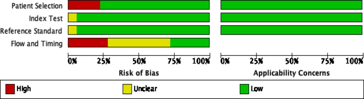
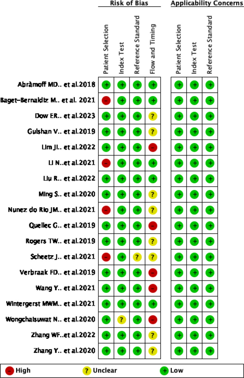
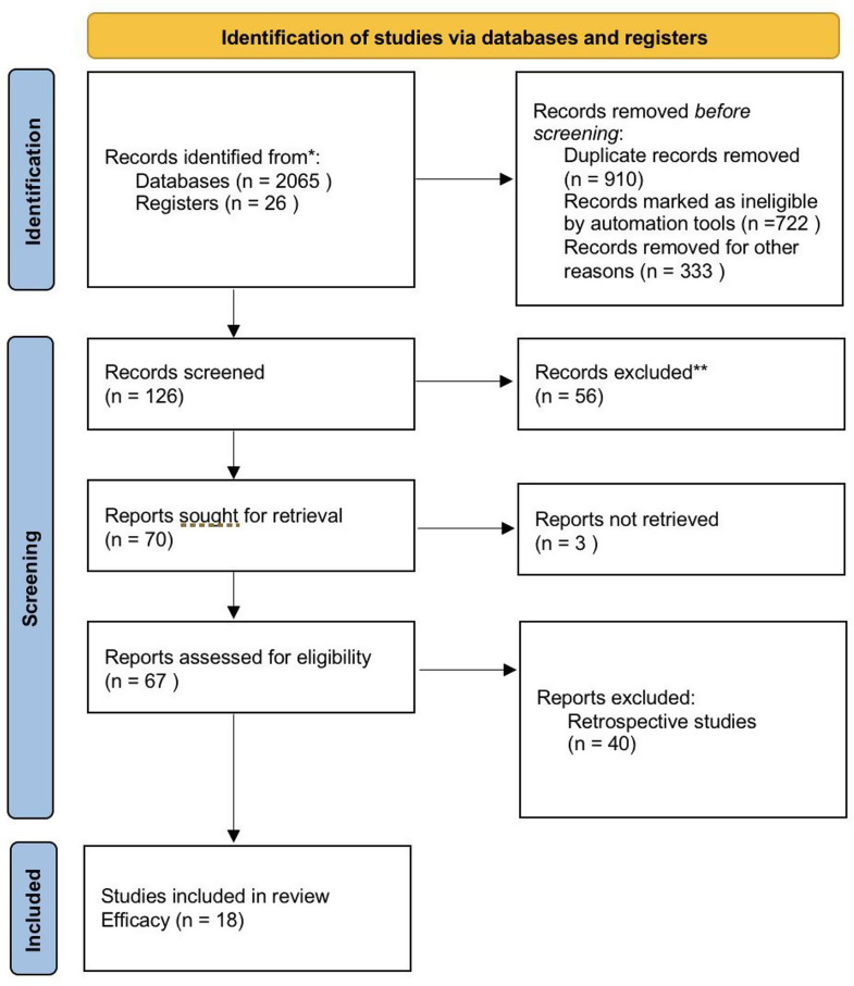
Methods: This systematic review was registered with PROSPERO (ID: CRD42024560750). Systematic searches were conducted in PubMed Medline, Cochrane Central, ScienceDirect, and Web of Science using keywords such as "diabetic retinopathy," "screening," and "artificial intelligence." Only studies published in English from 2019 to July 22, 2024, were considered. We also manually reviewed the reference lists of relevant reviews. Two independent reviewers assessed the risk of bias using the QUADAS-2 tool, resolving disagreements through discussion with the principal investigator. Meta-analysis was performed using MetaDiSc software (version 1.4). To calculate combined sensitivity, specificity, summary receiver operating characteristic (SROC) plots, forest plots, and subgroup analyses were performed according to clinician type (ophthalmologists vs. retina specialists) and imaging modality (fundus images vs. fundus images + OCT).
Results: 18 studies were included. Meta-analysis showed that AI systems demonstrated superior diagnostic performance compared to doctors, with the pooled sensitivity, specificity, diagnostic odds ratio, and Cochrane Q index of the AI being 0.877, 0.906, 0.94, and 153.79 accordingly. The Fagan nomogram analysis further confirmed the strong diagnostic value of AI. Subgroup analyses revealed that factors like imaging modality, and doctor expertise can influence diagnostic performance.
Conclusion: AI systems have demonstrated strong diagnostic performance in detecting diabetic retinopathy, with sensitivity and specificity comparable to or exceeding traditional clinicians.
期刊介绍:
International Journal of Retina and Vitreous focuses on the ophthalmic subspecialty of vitreoretinal disorders. The journal presents original articles on new approaches to diagnosis, outcomes of clinical trials, innovations in pharmacological therapy and surgical techniques, as well as basic science advances that impact clinical practice. Topical areas include, but are not limited to: -Imaging of the retina, choroid and vitreous -Innovations in optical coherence tomography (OCT) -Small-gauge vitrectomy, retinal detachment, chromovitrectomy -Electroretinography (ERG), microperimetry, other functional tests -Intraocular tumors -Retinal pharmacotherapy & drug delivery -Diabetic retinopathy & other vascular diseases -Age-related macular degeneration (AMD) & other macular entities

 求助内容:
求助内容: 应助结果提醒方式:


