IDEAL-IQ Magnetic Resonance Imaging Fat Fraction Quantification in Distinguishing Thymic Hyperplasia From Low-Risk Thymoma and Thymic Lymphoma in Adulthood: A Reliability and Efficacy Analysis.
IF 1.3 4区 医学Q4 RADIOLOGY, NUCLEAR MEDICINE & MEDICAL IMAGING
Jie Zhang, Xiu-Long Feng, Yu-Hui Ma, Jiang-Tao Lan, Shu-Mei Wang, Guang Yang, Yu-Chuan Hu, Guang-Bin Cui
{"title":"IDEAL-IQ Magnetic Resonance Imaging Fat Fraction Quantification in Distinguishing Thymic Hyperplasia From Low-Risk Thymoma and Thymic Lymphoma in Adulthood: A Reliability and Efficacy Analysis.","authors":"Jie Zhang, Xiu-Long Feng, Yu-Hui Ma, Jiang-Tao Lan, Shu-Mei Wang, Guang Yang, Yu-Chuan Hu, Guang-Bin Cui","doi":"10.1097/RCT.0000000000001688","DOIUrl":null,"url":null,"abstract":"<p><strong>Objectives: </strong>Detection of fat content in thymic lesions is essential to differentiate thymic hyperplasia from thymic tumors. This study assesses the reliability and efficacy of \"iterative decomposition of water and fat with echo asymmetry and least-squares estimation quantization\" IDEAL-IQ magnetic resonance sequence in distinguishing thymic hyperplasia from low-risk thymoma and thymic lymphoma in adulthood.</p><p><strong>Methods: </strong>Thirty patients with thymic hyperplasia, 28 low-risk thymomas, and 13 thymic lymphomas were respectively enrolled. All subjects underwent conventional thorax magnetic resonance imaging and IDEAL-IQ sequence. The fat fraction (FF mean and FF total ), signal intensity index, and R2* values of the lesions were compared for differences among 3 groups by the Mann-Whitney U and Kruskal-Wallis tests. Receiver operating characteristic curve analysis was performed to determine the differentiating efficacy.</p><p><strong>Results: </strong>Both FF mean and FF total values in patients with thymic hyperplasia are significantly higher than those in patients with low-risk thymoma and thymic lymphoma (FF mean : 26.41% vs 1.78% and 1.93%, FF total : 27.67% vs 2.21% and 2.44%; both P < 0.001), whereas there was no significant difference in these values between low-risk thymomas and thymic lymphomas (both P > 0.05). Similarly, signal intensity index and R2* values of thymic hyperplasia were significantly higher than those of patients with low-risk thymoma and thymic lymphoma ( P < 0.001). Receiver operating characteristic curve analysis showed that FF mean had an area under the curve of 0.998, with a cutoff of 4.78% yielding 95.12% sensitivity and 100% specificity, and FF total had an area under the curve of 0.994, with a cutoff of 8.57% yielding 97.56% sensitivity and 96.67% specificity in distinguishing thymic hyperplasia from tumors.</p><p><strong>Conclusions: </strong>IDEAL-IQ sequence provides accurate fat quantitative parameters and can differentiate thymic hyperplasia from thymic neoplasms with robust efficacy and reliability.</p>","PeriodicalId":15402,"journal":{"name":"Journal of Computer Assisted Tomography","volume":"49 3","pages":"431-439"},"PeriodicalIF":1.3000,"publicationDate":"2025-05-01","publicationTypes":"Journal Article","fieldsOfStudy":null,"isOpenAccess":false,"openAccessPdf":"https://www.ncbi.nlm.nih.gov/pmc/articles/PMC12071501/pdf/","citationCount":"0","resultStr":null,"platform":"Semanticscholar","paperid":null,"PeriodicalName":"Journal of Computer Assisted Tomography","FirstCategoryId":"3","ListUrlMain":"https://doi.org/10.1097/RCT.0000000000001688","RegionNum":4,"RegionCategory":"医学","ArticlePicture":[],"TitleCN":null,"AbstractTextCN":null,"PMCID":null,"EPubDate":"2024/11/18 0:00:00","PubModel":"Epub","JCR":"Q4","JCRName":"RADIOLOGY, NUCLEAR MEDICINE & MEDICAL IMAGING","Score":null,"Total":0}
引用次数: 0
Abstract
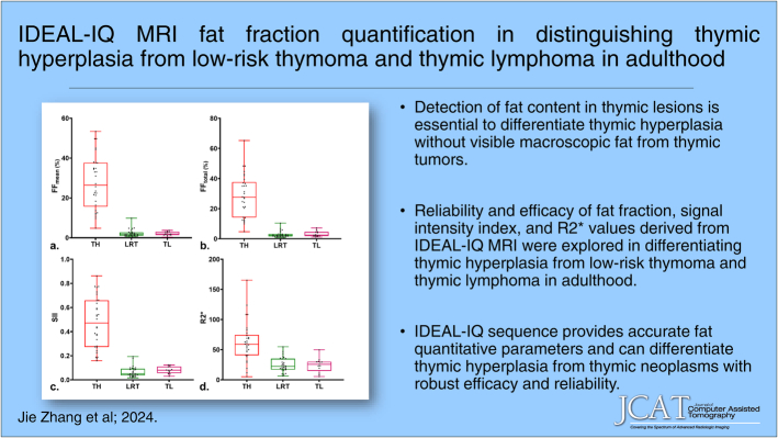
Objectives: Detection of fat content in thymic lesions is essential to differentiate thymic hyperplasia from thymic tumors. This study assesses the reliability and efficacy of "iterative decomposition of water and fat with echo asymmetry and least-squares estimation quantization" IDEAL-IQ magnetic resonance sequence in distinguishing thymic hyperplasia from low-risk thymoma and thymic lymphoma in adulthood.
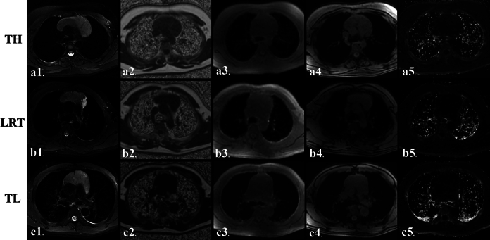
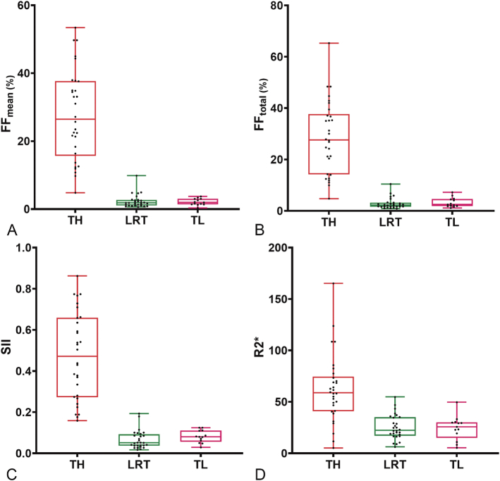
Methods: Thirty patients with thymic hyperplasia, 28 low-risk thymomas, and 13 thymic lymphomas were respectively enrolled. All subjects underwent conventional thorax magnetic resonance imaging and IDEAL-IQ sequence. The fat fraction (FF mean and FF total ), signal intensity index, and R2* values of the lesions were compared for differences among 3 groups by the Mann-Whitney U and Kruskal-Wallis tests. Receiver operating characteristic curve analysis was performed to determine the differentiating efficacy.
Results: Both FF mean and FF total values in patients with thymic hyperplasia are significantly higher than those in patients with low-risk thymoma and thymic lymphoma (FF mean : 26.41% vs 1.78% and 1.93%, FF total : 27.67% vs 2.21% and 2.44%; both P < 0.001), whereas there was no significant difference in these values between low-risk thymomas and thymic lymphomas (both P > 0.05). Similarly, signal intensity index and R2* values of thymic hyperplasia were significantly higher than those of patients with low-risk thymoma and thymic lymphoma ( P < 0.001). Receiver operating characteristic curve analysis showed that FF mean had an area under the curve of 0.998, with a cutoff of 4.78% yielding 95.12% sensitivity and 100% specificity, and FF total had an area under the curve of 0.994, with a cutoff of 8.57% yielding 97.56% sensitivity and 96.67% specificity in distinguishing thymic hyperplasia from tumors.
Conclusions: IDEAL-IQ sequence provides accurate fat quantitative parameters and can differentiate thymic hyperplasia from thymic neoplasms with robust efficacy and reliability.
目的:胸腺病变脂肪含量的检测是鉴别胸腺增生与胸腺肿瘤的重要依据。本研究评估了“回声不对称和最小二乘估计量化的反复分解水和脂肪”IDEAL-IQ磁共振序列在区分成年胸腺增生与低风险胸腺瘤和胸腺淋巴瘤中的可靠性和有效性。方法:选取胸腺增生患者30例,低危胸腺瘤患者28例,胸腺淋巴瘤患者13例。所有受试者均接受常规胸腔磁共振成像和IDEAL-IQ序列检查。采用Mann-Whitney U检验和Kruskal-Wallis检验比较3组间病变脂肪分数(FF均值和FF总量)、信号强度指数、R2*值的差异。采用受试者工作特征曲线分析,确定其鉴别疗效。结果:胸腺增生患者的FF均值和FF总值均显著高于低危胸腺瘤和胸腺淋巴瘤患者(FF均值:26.41% vs 1.78%和1.93%,FF总值:27.67% vs 2.21%和2.44%;P < 0.05)。胸腺增生的信号强度指数和R2*值也明显高于低危胸腺瘤和胸腺淋巴瘤(P)。结论:IDEAL-IQ序列提供了准确的脂肪定量参数,可以区分胸腺增生和胸腺肿瘤,具有较强的疗效和可靠性。
期刊介绍:
The mission of Journal of Computer Assisted Tomography is to showcase the latest clinical and research developments in CT, MR, and closely related diagnostic techniques. We encourage submission of both original research and review articles that have immediate or promissory clinical applications. Topics of special interest include: 1) functional MR and CT of the brain and body; 2) advanced/innovative MRI techniques (diffusion, perfusion, rapid scanning); and 3) advanced/innovative CT techniques (perfusion, multi-energy, dose-reduction, and processing).

 求助内容:
求助内容: 应助结果提醒方式:


