Macular thickness and vascular density assessment using optical coherence tomography and optical coherence tomography angiography imaging in iron ore mine personnel.
{"title":"Macular thickness and vascular density assessment using optical coherence tomography and optical coherence tomography angiography imaging in iron ore mine personnel.","authors":"Navid Faraji, Seyyed Pouria Tafti, Niloofar Khoshroo, Alireza Khoshrou, Elham Bakhtiari, Saeid Eslami, Nasser Shoeibi, Mohammad Reza Ansari Astaneh, Seyedeh Maryam Hosseini, Majid Abrishami, Hamid Reza Heidarzadeh, Parnian Arjmand, Mojtaba Abrishami","doi":"10.1186/s40942-025-00679-0","DOIUrl":null,"url":null,"abstract":"<p><strong>Background: </strong>To assess macular anatomical and vascular parameters in individuals working in iron ore mines using Optical Coherence Tomography (OCT) and Optical Coherence Tomography Angiography (OCTA) imaging to explore potential correlations between this occupational exposure and retinal changes.</p><p><strong>Methods: </strong>Individuals from the Sangan iron ore mine in Iran were included in a comparative cross-sectional observational study. An age-matched normal control group was selected from healthy participants employed at Mashhad University of Medical Sciences. Following thorough medical evaluations, participants underwent OCT and OCTA imaging. The macular thickness profile, vessel density (VD) of the superficial (SCP) and deep retinal capillary plexus (DCP), and the area of the foveal avascular zone (FAZ) were measured in our cases and compared with age-matched normal controls.</p><p><strong>Results: </strong>One hundred and one individuals, with an average age of 38.3 ± 5.59 years in the case group and 38.5 ± 5.59 years in the control group, were enrolled in the study. The difference in mean foveal thickness between cases (50.75 ± 9.13) and normal controls (50.38 ± 8.29) was not statistically significant (p = 0.758). Similarly, the mean VD in SCP and DCP for the case group (49.08 ± 2.20 and 49.32 ± 2.42, respectively) and the control group (49.45 ± 3.54 and 49.36 ± 3.97) did not show significant differences. Additionally, there were no significant changes (p-value > 0.05) in macular thickness and VD in other retinal regions when comparing the case and control groups.</p><p><strong>Conclusion: </strong>The research did not establish a significant association between occupational exposure in an iron ore mine and retinal structural changes or alterations in macular VD.</p>","PeriodicalId":14289,"journal":{"name":"International Journal of Retina and Vitreous","volume":"11 1","pages":"56"},"PeriodicalIF":2.4000,"publicationDate":"2025-05-09","publicationTypes":"Journal Article","fieldsOfStudy":null,"isOpenAccess":false,"openAccessPdf":"https://www.ncbi.nlm.nih.gov/pmc/articles/PMC12063320/pdf/","citationCount":"0","resultStr":null,"platform":"Semanticscholar","paperid":null,"PeriodicalName":"International Journal of Retina and Vitreous","FirstCategoryId":"1085","ListUrlMain":"https://doi.org/10.1186/s40942-025-00679-0","RegionNum":0,"RegionCategory":null,"ArticlePicture":[],"TitleCN":null,"AbstractTextCN":null,"PMCID":null,"EPubDate":"","PubModel":"","JCR":"Q2","JCRName":"OPHTHALMOLOGY","Score":null,"Total":0}
引用次数: 0
Abstract
Background: To assess macular anatomical and vascular parameters in individuals working in iron ore mines using Optical Coherence Tomography (OCT) and Optical Coherence Tomography Angiography (OCTA) imaging to explore potential correlations between this occupational exposure and retinal changes.
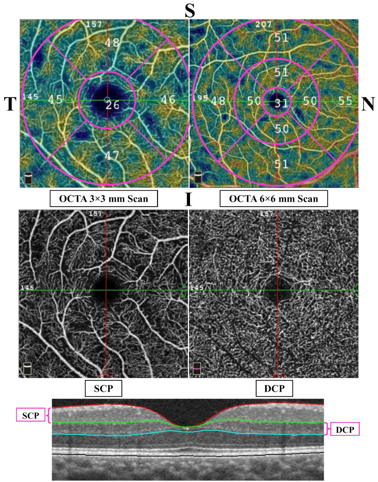
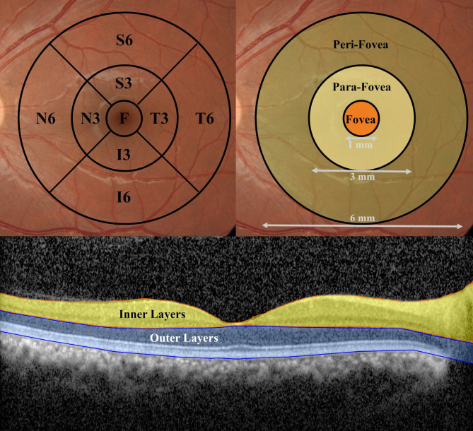
Methods: Individuals from the Sangan iron ore mine in Iran were included in a comparative cross-sectional observational study. An age-matched normal control group was selected from healthy participants employed at Mashhad University of Medical Sciences. Following thorough medical evaluations, participants underwent OCT and OCTA imaging. The macular thickness profile, vessel density (VD) of the superficial (SCP) and deep retinal capillary plexus (DCP), and the area of the foveal avascular zone (FAZ) were measured in our cases and compared with age-matched normal controls.
Results: One hundred and one individuals, with an average age of 38.3 ± 5.59 years in the case group and 38.5 ± 5.59 years in the control group, were enrolled in the study. The difference in mean foveal thickness between cases (50.75 ± 9.13) and normal controls (50.38 ± 8.29) was not statistically significant (p = 0.758). Similarly, the mean VD in SCP and DCP for the case group (49.08 ± 2.20 and 49.32 ± 2.42, respectively) and the control group (49.45 ± 3.54 and 49.36 ± 3.97) did not show significant differences. Additionally, there were no significant changes (p-value > 0.05) in macular thickness and VD in other retinal regions when comparing the case and control groups.
Conclusion: The research did not establish a significant association between occupational exposure in an iron ore mine and retinal structural changes or alterations in macular VD.
期刊介绍:
International Journal of Retina and Vitreous focuses on the ophthalmic subspecialty of vitreoretinal disorders. The journal presents original articles on new approaches to diagnosis, outcomes of clinical trials, innovations in pharmacological therapy and surgical techniques, as well as basic science advances that impact clinical practice. Topical areas include, but are not limited to: -Imaging of the retina, choroid and vitreous -Innovations in optical coherence tomography (OCT) -Small-gauge vitrectomy, retinal detachment, chromovitrectomy -Electroretinography (ERG), microperimetry, other functional tests -Intraocular tumors -Retinal pharmacotherapy & drug delivery -Diabetic retinopathy & other vascular diseases -Age-related macular degeneration (AMD) & other macular entities

 求助内容:
求助内容: 应助结果提醒方式:


