{"title":"Evaluation of the density of the midpalatal suture after maxillary expansion: a comparative observational study.","authors":"Gorkem Tekin, Yasin Caglar Kosar, Nesrin Saruhan Kose, Omur Dereci, Gizem Caliskan, Mehmet Ugurlu, Ayse Tugce Ozturk Kocak","doi":"10.1186/s13005-025-00508-7","DOIUrl":null,"url":null,"abstract":"<p><strong>Background: </strong>This study aimed to evaluate the effects of surgically assisted rapid maxillary expansion (SARME) and rapid maxillary expansion (RME) groups on midpalatal suture (MPS).</p><p><strong>Methods: </strong>CBCT records who underwent RME and SARME between 2013 and 2024 were included in the study. CBCT axial sections taken preoperatively (T0) and after a 3-month retention period (T1) were evaluated using the MPS. Fractal Analysis (FA) method using the ImageJ program and compared between the groups.</p><p><strong>Results: </strong>9 patients underwent SARME (%37.5) and 15 patients underwent RME (%62.5). FA values of the SARME and RME groups at T0 were found to be 1.02 ± 1.17 and 1.46 ± 0.09, respectively. FA values of the SARME and RME groups at T1 were found to be 0.98 ± 1.08 and 1.32 ± 0.08, respectively. The difference between T1 and T0 in the SARME and RME groups was 0.02 ± 0.09 and 0.34 ± 0.08, respectively. When FA differences were compared between the groups, no statistically significant difference was found. (p > 0.05) CONCLUSION: The potential effect of increasing retention time on the clinical recovery process has been clarified. In patients who underwent RME and SARME, after 3 months of retention, MPS density decreased compared to the initial density. The findings suggest that increasing the retention time in both RME and SARME groups for increased ossification. FA provides a useful method for evaluating skeletal effects of RME and SARME.</p>","PeriodicalId":12994,"journal":{"name":"Head & Face Medicine","volume":"21 1","pages":"30"},"PeriodicalIF":2.4000,"publicationDate":"2025-04-24","publicationTypes":"Journal Article","fieldsOfStudy":null,"isOpenAccess":false,"openAccessPdf":"https://www.ncbi.nlm.nih.gov/pmc/articles/PMC12020314/pdf/","citationCount":"0","resultStr":null,"platform":"Semanticscholar","paperid":null,"PeriodicalName":"Head & Face Medicine","FirstCategoryId":"3","ListUrlMain":"https://doi.org/10.1186/s13005-025-00508-7","RegionNum":2,"RegionCategory":"医学","ArticlePicture":[],"TitleCN":null,"AbstractTextCN":null,"PMCID":null,"EPubDate":"","PubModel":"","JCR":"Q2","JCRName":"DENTISTRY, ORAL SURGERY & MEDICINE","Score":null,"Total":0}
引用次数: 0
Abstract
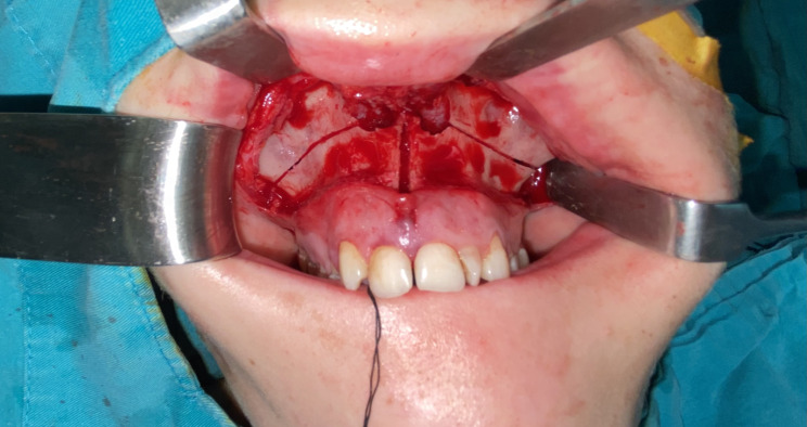
Background: This study aimed to evaluate the effects of surgically assisted rapid maxillary expansion (SARME) and rapid maxillary expansion (RME) groups on midpalatal suture (MPS).
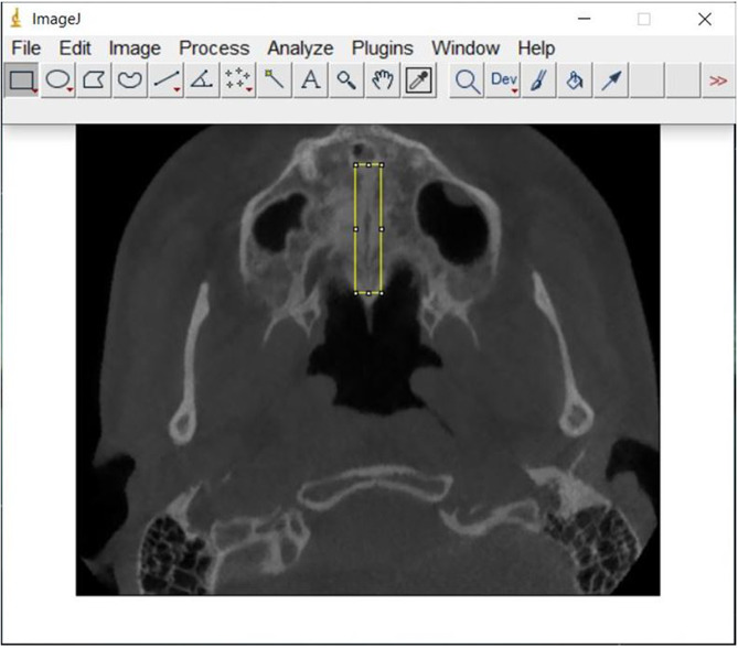
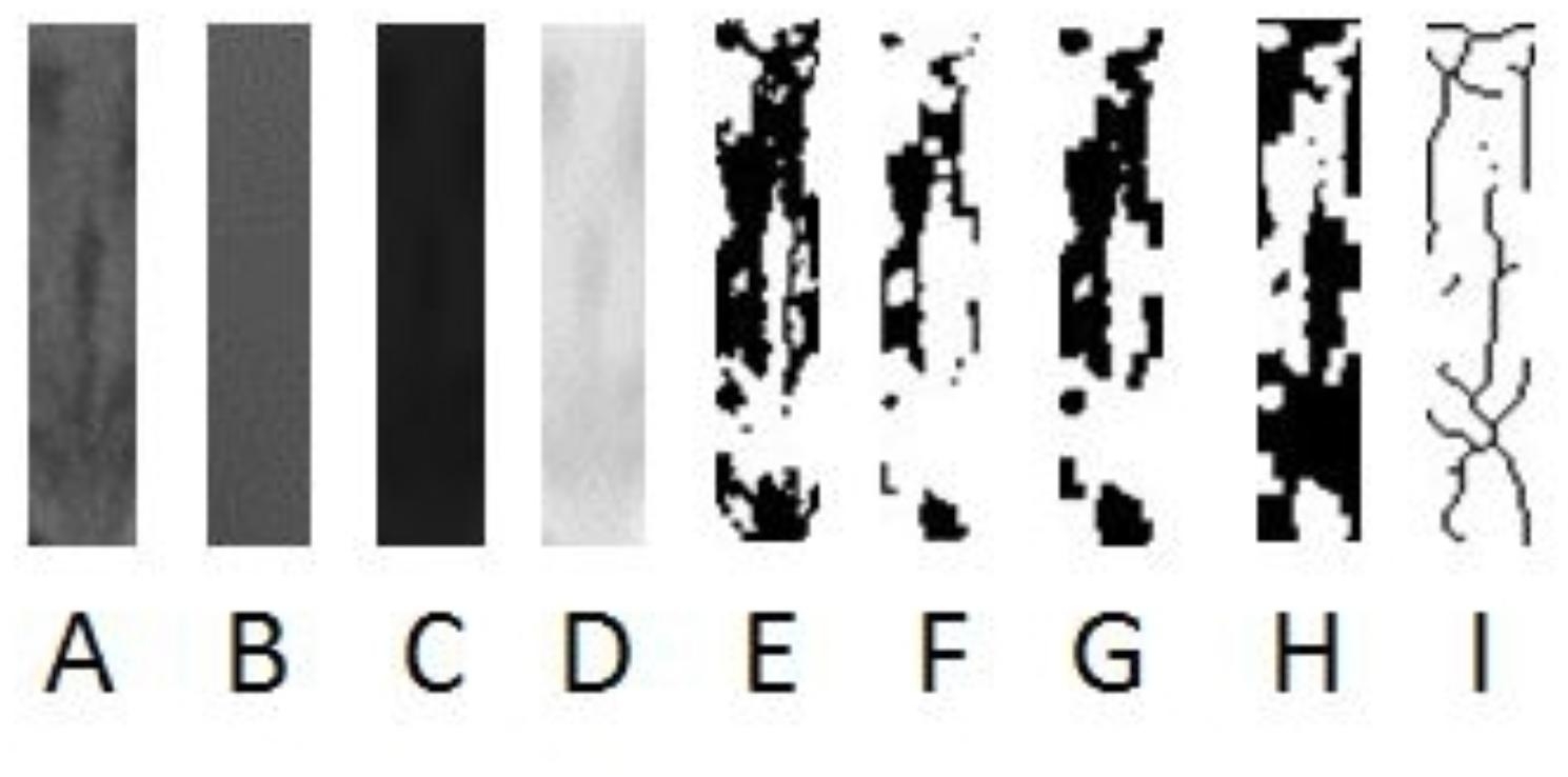
Methods: CBCT records who underwent RME and SARME between 2013 and 2024 were included in the study. CBCT axial sections taken preoperatively (T0) and after a 3-month retention period (T1) were evaluated using the MPS. Fractal Analysis (FA) method using the ImageJ program and compared between the groups.
Results: 9 patients underwent SARME (%37.5) and 15 patients underwent RME (%62.5). FA values of the SARME and RME groups at T0 were found to be 1.02 ± 1.17 and 1.46 ± 0.09, respectively. FA values of the SARME and RME groups at T1 were found to be 0.98 ± 1.08 and 1.32 ± 0.08, respectively. The difference between T1 and T0 in the SARME and RME groups was 0.02 ± 0.09 and 0.34 ± 0.08, respectively. When FA differences were compared between the groups, no statistically significant difference was found. (p > 0.05) CONCLUSION: The potential effect of increasing retention time on the clinical recovery process has been clarified. In patients who underwent RME and SARME, after 3 months of retention, MPS density decreased compared to the initial density. The findings suggest that increasing the retention time in both RME and SARME groups for increased ossification. FA provides a useful method for evaluating skeletal effects of RME and SARME.
期刊介绍:
Head & Face Medicine is a multidisciplinary open access journal that publishes basic and clinical research concerning all aspects of cranial, facial and oral conditions.
The journal covers all aspects of cranial, facial and oral diseases and their management. It has been designed as a multidisciplinary journal for clinicians and researchers involved in the diagnostic and therapeutic aspects of diseases which affect the human head and face. The journal is wide-ranging, covering the development, aetiology, epidemiology and therapy of head and face diseases to the basic science that underlies these diseases. Management of head and face diseases includes all aspects of surgical and non-surgical treatments including psychopharmacological therapies.
 求助内容:
求助内容: 应助结果提醒方式:


