Melanie Rieff, Fabian Holzberger, Oksana Lapina, Geir Ringstad, Lars Magnus Valnes, Bogna Warsza, Per Kristian Eide, Kent-André Mardal, Barbara Wohlmuth
{"title":"U-Net-Based Prediction of Cerebrospinal Fluid Distribution and Ventricular Reflux Grading.","authors":"Melanie Rieff, Fabian Holzberger, Oksana Lapina, Geir Ringstad, Lars Magnus Valnes, Bogna Warsza, Per Kristian Eide, Kent-André Mardal, Barbara Wohlmuth","doi":"10.1002/nbm.70029","DOIUrl":null,"url":null,"abstract":"<p><p>Previous work indicates evidence that cerebrospinal fluid (CSF) plays a crucial role in brain waste clearance processes and that altered flow patterns are associated with various diseases of the central nervous system. In this study, we investigate the potential of deep learning to predict the distribution in human brain of a gadolinium-based CSF contrast agent (tracer) administered intrathecal. For this, T1-weighted magnetic resonance imaging (MRI) scans taken at multiple time points before and after injection were utilized. We propose a U-net-based supervised learning model to predict pixel-wise signal increase at its peak after 24 h. Performance is evaluated based on different tracer distribution stages provided during training, including predictions from baseline scans taken before injection. Our findings show that training with imaging data from only the first 2-h postinjection yields tracer flow predictions comparable to models trained with additional later-stage scans. Validation against ventricular reflux gradings from neuroradiologists confirmed alignment with expert evaluations. These results demonstrate that deep learning-based methods for CSF flow prediction deserve more attention, as minimizing MR imaging without compromising clinical analysis could enhance efficiency, improve patient well-being and lower healthcare costs.</p>","PeriodicalId":19309,"journal":{"name":"NMR in Biomedicine","volume":"38 5","pages":"e70029"},"PeriodicalIF":2.7000,"publicationDate":"2025-05-01","publicationTypes":"Journal Article","fieldsOfStudy":null,"isOpenAccess":false,"openAccessPdf":"https://www.ncbi.nlm.nih.gov/pmc/articles/PMC11996590/pdf/","citationCount":"0","resultStr":null,"platform":"Semanticscholar","paperid":null,"PeriodicalName":"NMR in Biomedicine","FirstCategoryId":"3","ListUrlMain":"https://doi.org/10.1002/nbm.70029","RegionNum":4,"RegionCategory":"医学","ArticlePicture":[],"TitleCN":null,"AbstractTextCN":null,"PMCID":null,"EPubDate":"","PubModel":"","JCR":"Q2","JCRName":"BIOPHYSICS","Score":null,"Total":0}
引用次数: 0
Abstract
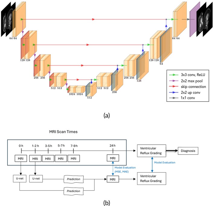
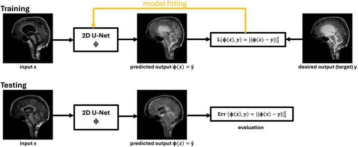
Previous work indicates evidence that cerebrospinal fluid (CSF) plays a crucial role in brain waste clearance processes and that altered flow patterns are associated with various diseases of the central nervous system. In this study, we investigate the potential of deep learning to predict the distribution in human brain of a gadolinium-based CSF contrast agent (tracer) administered intrathecal. For this, T1-weighted magnetic resonance imaging (MRI) scans taken at multiple time points before and after injection were utilized. We propose a U-net-based supervised learning model to predict pixel-wise signal increase at its peak after 24 h. Performance is evaluated based on different tracer distribution stages provided during training, including predictions from baseline scans taken before injection. Our findings show that training with imaging data from only the first 2-h postinjection yields tracer flow predictions comparable to models trained with additional later-stage scans. Validation against ventricular reflux gradings from neuroradiologists confirmed alignment with expert evaluations. These results demonstrate that deep learning-based methods for CSF flow prediction deserve more attention, as minimizing MR imaging without compromising clinical analysis could enhance efficiency, improve patient well-being and lower healthcare costs.
期刊介绍:
NMR in Biomedicine is a journal devoted to the publication of original full-length papers, rapid communications and review articles describing the development of magnetic resonance spectroscopy or imaging methods or their use to investigate physiological, biochemical, biophysical or medical problems. Topics for submitted papers should be in one of the following general categories: (a) development of methods and instrumentation for MR of biological systems; (b) studies of normal or diseased organs, tissues or cells; (c) diagnosis or treatment of disease. Reports may cover work on patients or healthy human subjects, in vivo animal experiments, studies of isolated organs or cultured cells, analysis of tissue extracts, NMR theory, experimental techniques, or instrumentation.

 求助内容:
求助内容: 应助结果提醒方式:


