Paula Andrea Sarmiento Riveros, Alejandro Jaramillo Quiceno, Rubén Darío Arias Pérez
{"title":"Patient-specific 3D tibial model: transforming meniscal allograft transplantation and surgical planning.","authors":"Paula Andrea Sarmiento Riveros, Alejandro Jaramillo Quiceno, Rubén Darío Arias Pérez","doi":"10.1186/s41205-025-00267-w","DOIUrl":null,"url":null,"abstract":"<p><strong>Background: </strong>Meniscal allograft transplantation (MAT) restores knee function by replacing a damaged or absent meniscus with a healthy allograft, helping to preserve joint stability, distribute the load, and reduce cartilage degeneration. However, traditional 2D imaging techniques fail to fully capture the knee's complex three-dimensional anatomy, making accurate surgical planning challenging. Computed Tomography (CT)-based 3D printing offers a patient-specific solution by generating anatomically precise tibial models, allowing for enhanced preoperative planning. This is particularly valuable in complex cases involving tibial osteotomy and anterior cruciate ligament (ACL) reconstruction, where precise tunnel positioning is critical to avoid tunnel convergence and ensure optimal graft integration.</p><p><strong>Case presentation: </strong>We present a case study and methodology demonstrating the generation and application of 3D-printed tibial models to assist in MAT, ACL reconstruction, and tibial osteotomy. High-resolution CT scans (slice thickness < 1 mm) were processed using D2P software to create a full-scale 3D model, which was printed using Hyper PLA filament. The 3D-printed model was provided to the tissue bank to optimize meniscal allograft selection and was integrated into preoperative planning to precisely determine tibial tunnel locations and angles, preventing overlap between MAT, ACL tunnels, and the osteotomy site. Intraoperatively, the model served as an accurate physical guide, facilitating osteophyte removal, guided tunnel drilling, and precise meniscal graft placement. Its use improved graft sizing accuracy minimized tunnel convergence, and allowed real-time intraoperative adjustments, which can improve surgical precision and decision-making.</p><p><strong>Conclusions: </strong>The integration of patient-specific 3D-printed models into surgical planning and execution may improve accuracy and efficiency in complex MAT procedures that also involve tibial osteotomy and ACL reconstruction. These models offer detailed anatomical reference points that facilitate more precise graft selection, tunnel placement, and intraoperative decision-making. However, further studies are needed to validate their dimensional accuracy, evaluate clinical outcomes in larger cohorts, and determine their feasibility for routine use in orthopedic practice.</p>","PeriodicalId":72036,"journal":{"name":"3D printing in medicine","volume":"11 1","pages":"20"},"PeriodicalIF":3.1000,"publicationDate":"2025-05-06","publicationTypes":"Journal Article","fieldsOfStudy":null,"isOpenAccess":false,"openAccessPdf":"https://www.ncbi.nlm.nih.gov/pmc/articles/PMC12054210/pdf/","citationCount":"0","resultStr":null,"platform":"Semanticscholar","paperid":null,"PeriodicalName":"3D printing in medicine","FirstCategoryId":"1085","ListUrlMain":"https://doi.org/10.1186/s41205-025-00267-w","RegionNum":0,"RegionCategory":null,"ArticlePicture":[],"TitleCN":null,"AbstractTextCN":null,"PMCID":null,"EPubDate":"","PubModel":"","JCR":"Q1","JCRName":"RADIOLOGY, NUCLEAR MEDICINE & MEDICAL IMAGING","Score":null,"Total":0}
引用次数: 0
Abstract
Background: Meniscal allograft transplantation (MAT) restores knee function by replacing a damaged or absent meniscus with a healthy allograft, helping to preserve joint stability, distribute the load, and reduce cartilage degeneration. However, traditional 2D imaging techniques fail to fully capture the knee's complex three-dimensional anatomy, making accurate surgical planning challenging. Computed Tomography (CT)-based 3D printing offers a patient-specific solution by generating anatomically precise tibial models, allowing for enhanced preoperative planning. This is particularly valuable in complex cases involving tibial osteotomy and anterior cruciate ligament (ACL) reconstruction, where precise tunnel positioning is critical to avoid tunnel convergence and ensure optimal graft integration.
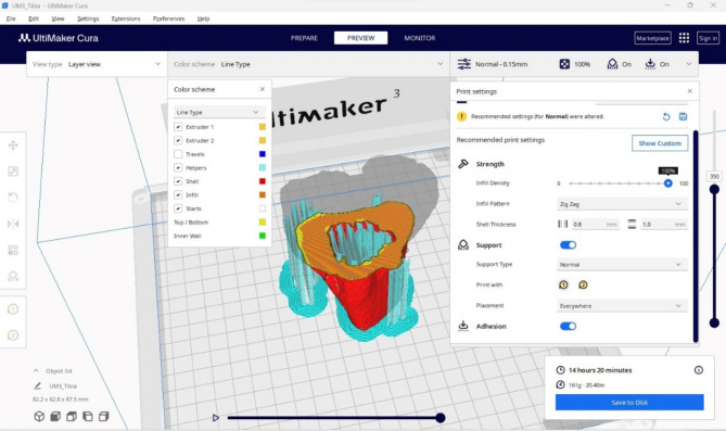
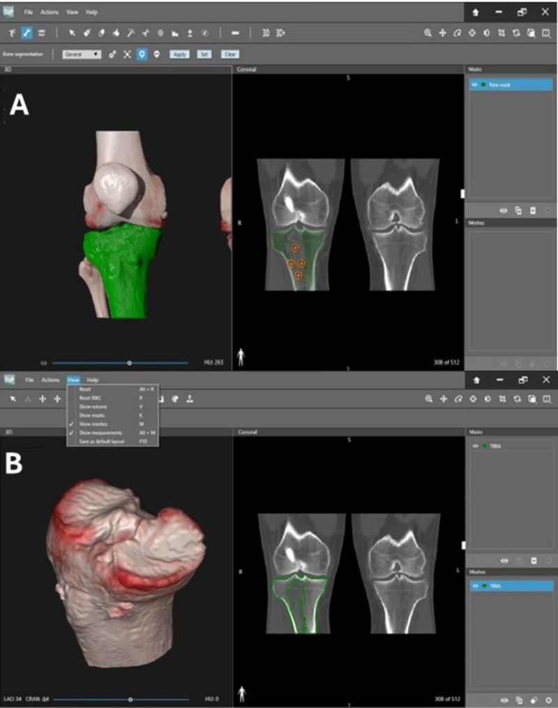
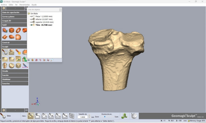
Case presentation: We present a case study and methodology demonstrating the generation and application of 3D-printed tibial models to assist in MAT, ACL reconstruction, and tibial osteotomy. High-resolution CT scans (slice thickness < 1 mm) were processed using D2P software to create a full-scale 3D model, which was printed using Hyper PLA filament. The 3D-printed model was provided to the tissue bank to optimize meniscal allograft selection and was integrated into preoperative planning to precisely determine tibial tunnel locations and angles, preventing overlap between MAT, ACL tunnels, and the osteotomy site. Intraoperatively, the model served as an accurate physical guide, facilitating osteophyte removal, guided tunnel drilling, and precise meniscal graft placement. Its use improved graft sizing accuracy minimized tunnel convergence, and allowed real-time intraoperative adjustments, which can improve surgical precision and decision-making.
Conclusions: The integration of patient-specific 3D-printed models into surgical planning and execution may improve accuracy and efficiency in complex MAT procedures that also involve tibial osteotomy and ACL reconstruction. These models offer detailed anatomical reference points that facilitate more precise graft selection, tunnel placement, and intraoperative decision-making. However, further studies are needed to validate their dimensional accuracy, evaluate clinical outcomes in larger cohorts, and determine their feasibility for routine use in orthopedic practice.

 求助内容:
求助内容: 应助结果提醒方式:


