Yahya Alperen Bayraktar, Mehmet Ali Eryılmaz, Mehmet Eşref Ulutaş, Alpaslan Şahin, Gürcan Şimşek, Şerife Yüksekkaya
{"title":"Assessment of the Scolicidal Effect of Bile: An <i>Ex Vivo</i> Study.","authors":"Yahya Alperen Bayraktar, Mehmet Ali Eryılmaz, Mehmet Eşref Ulutaş, Alpaslan Şahin, Gürcan Şimşek, Şerife Yüksekkaya","doi":"10.18502/ijpa.v20i1.18108","DOIUrl":null,"url":null,"abstract":"<p><strong>Background: </strong>Approximately 25% of hepatic hydatid cysts rupture into the biliary tract. The precise effect of bile within the cyst on protoscoleces remains unclear. We aimed to elucidate the effect of bile on protoscoleces.</p><p><strong>Methods: </strong>The contents of hydatid cysts from the livers of three sheep were aspirated under sterile conditions. The aspirated contents were divided into 50 separate Eppendorf tubes (5 cc). Samples from each tube were stained with 0.1% eosin Y. Pink stained protoscoleces were considered dead under light microscopy (×100). A total of 100 protoscoleces were counted in each sample, and the number of live and dead protoscoleces was recorded. The tubes were randomly divided into five groups. Group 1 served as the control, Group 2 received normal saline (NS), Group 3, received hypertonic saline, Group 4 received bile, and Group 4 received diluted bile. The number of live and dead protoscoleces was recorded at the end of the first and second hours.</p><p><strong>Results: </strong>Compared to the initial count of live protoscoleces, the number of live protoscoleces increased at hours 1 and 2 in Groups 2 and 4. No live protoscoleces remained at hours 1 and 2 in Group 3. There was no significant change in Group 5. When compared to the control group, a significant increase in viability was observed only in Group 4 (<i>P</i>=0.001).</p><p><strong>Conclusion: </strong>Bile of sheep does not exhibit scolicidal effects; rather, it positively affects protoscoleces by increasing viability.</p>","PeriodicalId":14669,"journal":{"name":"Iranian Journal of Parasitology","volume":"20 1","pages":"83-90"},"PeriodicalIF":0.9000,"publicationDate":"2025-01-01","publicationTypes":"Journal Article","fieldsOfStudy":null,"isOpenAccess":false,"openAccessPdf":"https://www.ncbi.nlm.nih.gov/pmc/articles/PMC11978201/pdf/","citationCount":"0","resultStr":null,"platform":"Semanticscholar","paperid":null,"PeriodicalName":"Iranian Journal of Parasitology","FirstCategoryId":"3","ListUrlMain":"https://doi.org/10.18502/ijpa.v20i1.18108","RegionNum":4,"RegionCategory":"医学","ArticlePicture":[],"TitleCN":null,"AbstractTextCN":null,"PMCID":null,"EPubDate":"","PubModel":"","JCR":"Q4","JCRName":"PARASITOLOGY","Score":null,"Total":0}
引用次数: 0
Abstract
Background: Approximately 25% of hepatic hydatid cysts rupture into the biliary tract. The precise effect of bile within the cyst on protoscoleces remains unclear. We aimed to elucidate the effect of bile on protoscoleces.
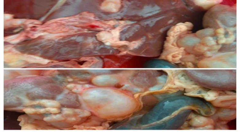
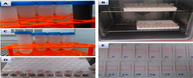
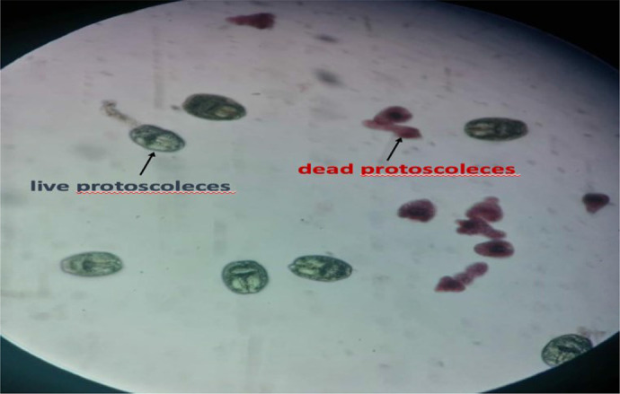
Methods: The contents of hydatid cysts from the livers of three sheep were aspirated under sterile conditions. The aspirated contents were divided into 50 separate Eppendorf tubes (5 cc). Samples from each tube were stained with 0.1% eosin Y. Pink stained protoscoleces were considered dead under light microscopy (×100). A total of 100 protoscoleces were counted in each sample, and the number of live and dead protoscoleces was recorded. The tubes were randomly divided into five groups. Group 1 served as the control, Group 2 received normal saline (NS), Group 3, received hypertonic saline, Group 4 received bile, and Group 4 received diluted bile. The number of live and dead protoscoleces was recorded at the end of the first and second hours.
Results: Compared to the initial count of live protoscoleces, the number of live protoscoleces increased at hours 1 and 2 in Groups 2 and 4. No live protoscoleces remained at hours 1 and 2 in Group 3. There was no significant change in Group 5. When compared to the control group, a significant increase in viability was observed only in Group 4 (P=0.001).
Conclusion: Bile of sheep does not exhibit scolicidal effects; rather, it positively affects protoscoleces by increasing viability.
期刊介绍:
Iranian Journal of Parasitology (IJP) is the official publication of Iranian Society of Parasitology (ISP) launched in 2006. The society was inaugurated in 1994 and pursues the improvement of the knowledge on the parasites and parasitic diseases, exchange of scientific knowledge with foreign societies, publicity activities, and consultation on the parasitic diseases, and intimate relationship among society members.
The main aims of the Journal are: contribution to the field of Parasitology, including all aspects of parasites and parasitic diseases (medical and veterinary) and related fields such as Entomology which may be submitted by scientists from Iran and all over the world.
 求助内容:
求助内容: 应助结果提醒方式:


