{"title":"Technical report: Calculation and interpretation of corneal transference maps.","authors":"Tanya Evans, Jos J Rozema","doi":"10.1097/OPX.0000000000002248","DOIUrl":null,"url":null,"abstract":"<p><strong>Significance: </strong>Transference matrices are interesting tools for optical analysis of an eye alongside ray tracing. To explore possible interpretations of the ABCD entries of the transference, this article applies them to the corneal surfaces to find a surprising link to a corneal biomechanics parameter that may help with keratoconus detection.</p><p><strong>Purpose: </strong>The transference matrix used in linear optics has four entries, dilation A, disjugacy B, divergence C, and divarication D. Entry C is the negative of the power of the system. However, the remaining three entries are somewhat difficult to relate to. This work explores how the four fundamental properties of the corneal transference relate to familiar corneal variables such as radii of curvature, thickness, surface powers, and total refractive power.</p><p><strong>Methods: </strong>The cornea is treated as a thick lens, and a transference is obtained for the cornea at approximately 12,000 points, as well as point-by-point corneal maps of A, B, C, and D are obtained based on Scheimpflug tomography data (Pentacam HR, Wetzlar, Germany). The trace of the transference is also obtained.</p><p><strong>Results: </strong>The four corneal maps of A, B, C, and D resemble familiar clinical maps, albeit at different scales. Pachymetry is represented by B, and total corneal power is represented by C, and A represents a new variable, resembling the corneal contribution to stress (CCS), a new variable used in detecting early keratoconus. D seems to represent a CCS-like variable applied to the posterior corneal surface. In keratoconus, the trace appears as a ring-shaped pattern around the cone.</p><p><strong>Conclusions: </strong>The A, B, C, and D maps relate information from known clinical maps such as pachymetry, corneal power, and CCS. The trace of the transference provides a new corneal map representing the combination of CCS and a related posterior parameter that may be useful in the detection and follow-up of keratoconus.</p>","PeriodicalId":19649,"journal":{"name":"Optometry and Vision Science","volume":"102 4","pages":"228-234"},"PeriodicalIF":1.8000,"publicationDate":"2025-04-01","publicationTypes":"Journal Article","fieldsOfStudy":null,"isOpenAccess":false,"openAccessPdf":"https://www.ncbi.nlm.nih.gov/pmc/articles/PMC12147723/pdf/","citationCount":"0","resultStr":null,"platform":"Semanticscholar","paperid":null,"PeriodicalName":"Optometry and Vision Science","FirstCategoryId":"3","ListUrlMain":"https://doi.org/10.1097/OPX.0000000000002248","RegionNum":4,"RegionCategory":"医学","ArticlePicture":[],"TitleCN":null,"AbstractTextCN":null,"PMCID":null,"EPubDate":"","PubModel":"","JCR":"Q3","JCRName":"OPHTHALMOLOGY","Score":null,"Total":0}
引用次数: 0
Abstract
Significance: Transference matrices are interesting tools for optical analysis of an eye alongside ray tracing. To explore possible interpretations of the ABCD entries of the transference, this article applies them to the corneal surfaces to find a surprising link to a corneal biomechanics parameter that may help with keratoconus detection.
Purpose: The transference matrix used in linear optics has four entries, dilation A, disjugacy B, divergence C, and divarication D. Entry C is the negative of the power of the system. However, the remaining three entries are somewhat difficult to relate to. This work explores how the four fundamental properties of the corneal transference relate to familiar corneal variables such as radii of curvature, thickness, surface powers, and total refractive power.
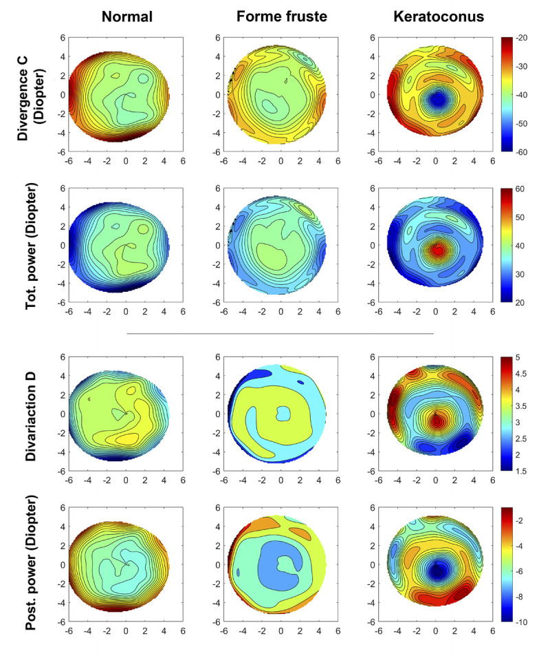
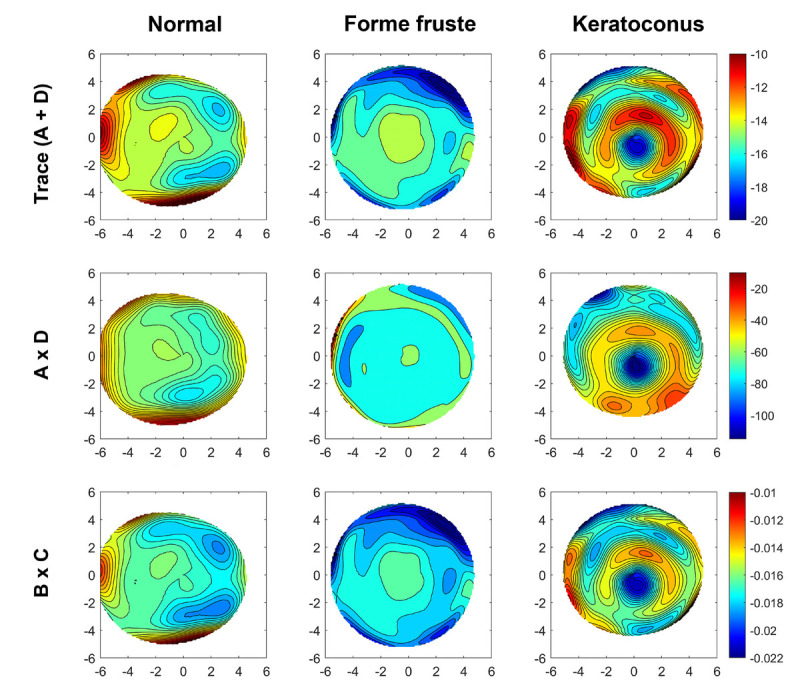
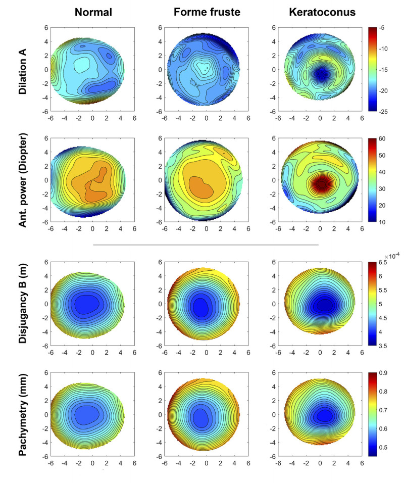
Methods: The cornea is treated as a thick lens, and a transference is obtained for the cornea at approximately 12,000 points, as well as point-by-point corneal maps of A, B, C, and D are obtained based on Scheimpflug tomography data (Pentacam HR, Wetzlar, Germany). The trace of the transference is also obtained.
Results: The four corneal maps of A, B, C, and D resemble familiar clinical maps, albeit at different scales. Pachymetry is represented by B, and total corneal power is represented by C, and A represents a new variable, resembling the corneal contribution to stress (CCS), a new variable used in detecting early keratoconus. D seems to represent a CCS-like variable applied to the posterior corneal surface. In keratoconus, the trace appears as a ring-shaped pattern around the cone.
Conclusions: The A, B, C, and D maps relate information from known clinical maps such as pachymetry, corneal power, and CCS. The trace of the transference provides a new corneal map representing the combination of CCS and a related posterior parameter that may be useful in the detection and follow-up of keratoconus.
期刊介绍:
Optometry and Vision Science is the monthly peer-reviewed scientific publication of the American Academy of Optometry, publishing original research since 1924. Optometry and Vision Science is an internationally recognized source for education and information on current discoveries in optometry, physiological optics, vision science, and related fields. The journal considers original contributions that advance clinical practice, vision science, and public health. Authors should remember that the journal reaches readers worldwide and their submissions should be relevant and of interest to a broad audience. Topical priorities include, but are not limited to: clinical and laboratory research, evidence-based reviews, contact lenses, ocular growth and refractive error development, eye movements, visual function and perception, biology of the eye and ocular disease, epidemiology and public health, biomedical optics and instrumentation, novel and important clinical observations and treatments, and optometric education.
 求助内容:
求助内容: 应助结果提醒方式:


