Ahmed Ibrahim Basiony, Sameh Mohamed Elgouhary, Hadeer Elbasuony Mohamed, Enas Sobhy Zahran
{"title":"Assessment of retinal microvascular changes in patients with systemic lupus erythematosus using optical coherence tomography angiography.","authors":"Ahmed Ibrahim Basiony, Sameh Mohamed Elgouhary, Hadeer Elbasuony Mohamed, Enas Sobhy Zahran","doi":"10.1186/s40942-025-00677-2","DOIUrl":null,"url":null,"abstract":"<p><strong>Background: </strong>It is evident that the physiopathological pathways of ocular and renal microvascular tissues in patients with systemic lupus are similar. Previously, this was confirmed by employing traditional fundus examination, optical coherence tomography, and high-resolution color electroretinography. Recent years have seen the development of Optical Coherence Tomography Angiography (OCTA) as a non-intrusive procedure that can be employed to image the microvasculature of the retina and choroid.</p><p><strong>Objective: </strong>The aim of this study is to assess the correlation between renal functional and histologic features with the retinal microvasculature alterations in systemic lupus patients through OCTA analysis.</p><p><strong>Patients and methods: </strong>This case-control study enrolled thirty-six eyes from 18 lupus nephritis (LN) patients, thirty-six eyes from 18 systemic lupus erythematosus (SLE) patients, and thirty eyes from 15 healthy controls. An ophthalmological evaluation, including history, examination, and investigations, was conducted using OCTA for all participants. Prior to ocular examination and investigation, all SLE patients underwent a rheumatological evaluation, encompassing disease-related clinical and laboratory assessments. Specimen retrieval and renal biopsy examinations were also performed, categorizing them into lupus and lupus nephritis patients.</p><p><strong>Results: </strong>Regarding central foveal thickness (CFT) and parafoveal thickness (PFT), there were no significant differences compared to healthy subjects. A comparison of the foveal avascular zone area (FAZ-A) among the three groups revealed a significant increase in both patient groups compared to healthy controls. Whole superficial capillary plexus (SCP) vascular density (VD) in the parafoveal and foveal regions showed a significant reduction in both SLE patient groups compared to healthy controls (HC). Specifically, SCP values were 42.65 ± 2.23% in the SLE with nephritis group, 44.88 ± 2.09% in the SLE without nephritis group, and 49.10 ± 3.12% in the healthy control group. SCP parafoveal VD values were 40.77 ± 3.27% in SLE with nephritis, 47.19 ± 2.63% in SLE without nephritis, and 50.98 ± 4.80% in healthy controls. SCP foveal VD was 18.96 ± 3.43% in SLE with nephritis, 21.61 ± 4.00% in SLE without nephritis, and 24.16 ± 2.69% in healthy controls. The whole deep capillary plexus (DCP), parafoveal, and foveal VD were significantly reduced in the SLE with nephritis group but showed only marginal differences in the SLE without nephritis group compared to healthy controls, as DCP values were 48.04 ± 3.93% in SLE with nephritis, 53.63 ± 2.19% in SLE without nephritis, and 54.88 ± 3.57% in healthy controls. DCP parafoveal VD was 54.56 ± 2.37% in SLE with nephritis, 56.93 ± 1.90% in SLE without nephritis, and 57.39 ± 5.99% in healthy controls. DCP foveal VD was 34.42 ± 3.12% in SLE with nephritis, 41.96 ± 3.19% in SLE without nephritis, and 42.55 ± 7.74% in healthy controls.</p><p><strong>Conclusion: </strong>OCT angiography has a considerable role in the detection of the early changes of the retinal vascular plexus in patients with SLE, especially those with lupus nephritis, even before the development of retinopathy.</p>","PeriodicalId":14289,"journal":{"name":"International Journal of Retina and Vitreous","volume":"11 1","pages":"55"},"PeriodicalIF":2.4000,"publicationDate":"2025-05-08","publicationTypes":"Journal Article","fieldsOfStudy":null,"isOpenAccess":false,"openAccessPdf":"https://www.ncbi.nlm.nih.gov/pmc/articles/PMC12060311/pdf/","citationCount":"0","resultStr":null,"platform":"Semanticscholar","paperid":null,"PeriodicalName":"International Journal of Retina and Vitreous","FirstCategoryId":"1085","ListUrlMain":"https://doi.org/10.1186/s40942-025-00677-2","RegionNum":0,"RegionCategory":null,"ArticlePicture":[],"TitleCN":null,"AbstractTextCN":null,"PMCID":null,"EPubDate":"","PubModel":"","JCR":"Q2","JCRName":"OPHTHALMOLOGY","Score":null,"Total":0}
引用次数: 0
Abstract
Background: It is evident that the physiopathological pathways of ocular and renal microvascular tissues in patients with systemic lupus are similar. Previously, this was confirmed by employing traditional fundus examination, optical coherence tomography, and high-resolution color electroretinography. Recent years have seen the development of Optical Coherence Tomography Angiography (OCTA) as a non-intrusive procedure that can be employed to image the microvasculature of the retina and choroid.
Objective: The aim of this study is to assess the correlation between renal functional and histologic features with the retinal microvasculature alterations in systemic lupus patients through OCTA analysis.
Patients and methods: This case-control study enrolled thirty-six eyes from 18 lupus nephritis (LN) patients, thirty-six eyes from 18 systemic lupus erythematosus (SLE) patients, and thirty eyes from 15 healthy controls. An ophthalmological evaluation, including history, examination, and investigations, was conducted using OCTA for all participants. Prior to ocular examination and investigation, all SLE patients underwent a rheumatological evaluation, encompassing disease-related clinical and laboratory assessments. Specimen retrieval and renal biopsy examinations were also performed, categorizing them into lupus and lupus nephritis patients.
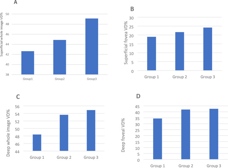
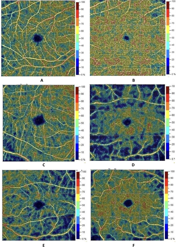
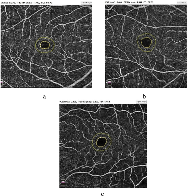
Results: Regarding central foveal thickness (CFT) and parafoveal thickness (PFT), there were no significant differences compared to healthy subjects. A comparison of the foveal avascular zone area (FAZ-A) among the three groups revealed a significant increase in both patient groups compared to healthy controls. Whole superficial capillary plexus (SCP) vascular density (VD) in the parafoveal and foveal regions showed a significant reduction in both SLE patient groups compared to healthy controls (HC). Specifically, SCP values were 42.65 ± 2.23% in the SLE with nephritis group, 44.88 ± 2.09% in the SLE without nephritis group, and 49.10 ± 3.12% in the healthy control group. SCP parafoveal VD values were 40.77 ± 3.27% in SLE with nephritis, 47.19 ± 2.63% in SLE without nephritis, and 50.98 ± 4.80% in healthy controls. SCP foveal VD was 18.96 ± 3.43% in SLE with nephritis, 21.61 ± 4.00% in SLE without nephritis, and 24.16 ± 2.69% in healthy controls. The whole deep capillary plexus (DCP), parafoveal, and foveal VD were significantly reduced in the SLE with nephritis group but showed only marginal differences in the SLE without nephritis group compared to healthy controls, as DCP values were 48.04 ± 3.93% in SLE with nephritis, 53.63 ± 2.19% in SLE without nephritis, and 54.88 ± 3.57% in healthy controls. DCP parafoveal VD was 54.56 ± 2.37% in SLE with nephritis, 56.93 ± 1.90% in SLE without nephritis, and 57.39 ± 5.99% in healthy controls. DCP foveal VD was 34.42 ± 3.12% in SLE with nephritis, 41.96 ± 3.19% in SLE without nephritis, and 42.55 ± 7.74% in healthy controls.
Conclusion: OCT angiography has a considerable role in the detection of the early changes of the retinal vascular plexus in patients with SLE, especially those with lupus nephritis, even before the development of retinopathy.
期刊介绍:
International Journal of Retina and Vitreous focuses on the ophthalmic subspecialty of vitreoretinal disorders. The journal presents original articles on new approaches to diagnosis, outcomes of clinical trials, innovations in pharmacological therapy and surgical techniques, as well as basic science advances that impact clinical practice. Topical areas include, but are not limited to: -Imaging of the retina, choroid and vitreous -Innovations in optical coherence tomography (OCT) -Small-gauge vitrectomy, retinal detachment, chromovitrectomy -Electroretinography (ERG), microperimetry, other functional tests -Intraocular tumors -Retinal pharmacotherapy & drug delivery -Diabetic retinopathy & other vascular diseases -Age-related macular degeneration (AMD) & other macular entities

 求助内容:
求助内容: 应助结果提醒方式:


