Eun Kyung Khil, Jang Gyu Cha, Sung Jae Kim, Yu Sung Yoon
{"title":"Quantitative T2 Mapping Analysis With MRI of Talar Cartilage in Ankle Trauma: A Study Based on Lauge-Hansen Classification and Anatomical Locations.","authors":"Eun Kyung Khil, Jang Gyu Cha, Sung Jae Kim, Yu Sung Yoon","doi":"10.3348/kjr.2024.0773","DOIUrl":null,"url":null,"abstract":"<p><strong>Objective: </strong>This study aimed to quantitatively assess abnormalities in the talar dome cartilage using MRI T2 mapping, with additional analyses based on the Lauge-Hansen (LH) classification and anatomical locations.</p><p><strong>Materials and methods: </strong>This retrospective study analyzed 78 patients who underwent ankle MRI with T2 mapping for acute ankle trauma between January 2021 and October 2022. Patients were classified into the supination (S) and pronation (P) groups based on the LH classification, and then divided into subgroups based on posterior malleolus (PM) involvement. The T2 values for the talar cartilage were quantitatively measured in six anatomical regions defined by the combination of medial vs. lateral and anterior vs. central vs. posterior. The T2 mapping values in each region of the talus were compared between the S and P groups and between the PM and non-PM injury groups using <i>t</i>-tests. The T2 values were also compared between the medial and lateral sides within each group.</p><p><strong>Results: </strong>Among the 78 patients (mean age, 38.62 ± 14.82 years; 47 male), 53 and 25 were in the S and P groups, respectively, and 53 patients showed PM involvement. In comparison with the P group, the S group exhibited higher T2 values in the medial portion (61.27 ± 8.30 vs. 54.03 ± 6.96; <i>P</i> < 0.001) and lower T2 values in the lateral talus (54.95 ± 8.47 vs. 64.15 ± 7.31; <i>P</i> < 0.001). The PM injury group showed higher T2 values in the posterior region than the non-PM injury group (<i>P</i> ≤ 0.011). Within the PM injury group, T2 values were higher in the anteromedial and posterolateral regions than on the opposite sides (<i>P</i> = 0.037 and 0.011, respectively).</p><p><strong>Conclusion: </strong>MRI T2 values demonstrated significant regional variations in the talar dome cartilage in acute ankle trauma, and the T2 values may reflect different ankle trauma mechanisms and PM involvement. Thus, T2 mapping can facilitate evaluation of talar cartilage alterations.</p>","PeriodicalId":17881,"journal":{"name":"Korean Journal of Radiology","volume":"26 5","pages":"435-445"},"PeriodicalIF":5.3000,"publicationDate":"2025-05-01","publicationTypes":"Journal Article","fieldsOfStudy":null,"isOpenAccess":false,"openAccessPdf":"https://www.ncbi.nlm.nih.gov/pmc/articles/PMC12055265/pdf/","citationCount":"0","resultStr":null,"platform":"Semanticscholar","paperid":null,"PeriodicalName":"Korean Journal of Radiology","FirstCategoryId":"3","ListUrlMain":"https://doi.org/10.3348/kjr.2024.0773","RegionNum":2,"RegionCategory":"医学","ArticlePicture":[],"TitleCN":null,"AbstractTextCN":null,"PMCID":null,"EPubDate":"","PubModel":"","JCR":"Q1","JCRName":"RADIOLOGY, NUCLEAR MEDICINE & MEDICAL IMAGING","Score":null,"Total":0}
引用次数: 0
Abstract
Objective: This study aimed to quantitatively assess abnormalities in the talar dome cartilage using MRI T2 mapping, with additional analyses based on the Lauge-Hansen (LH) classification and anatomical locations.
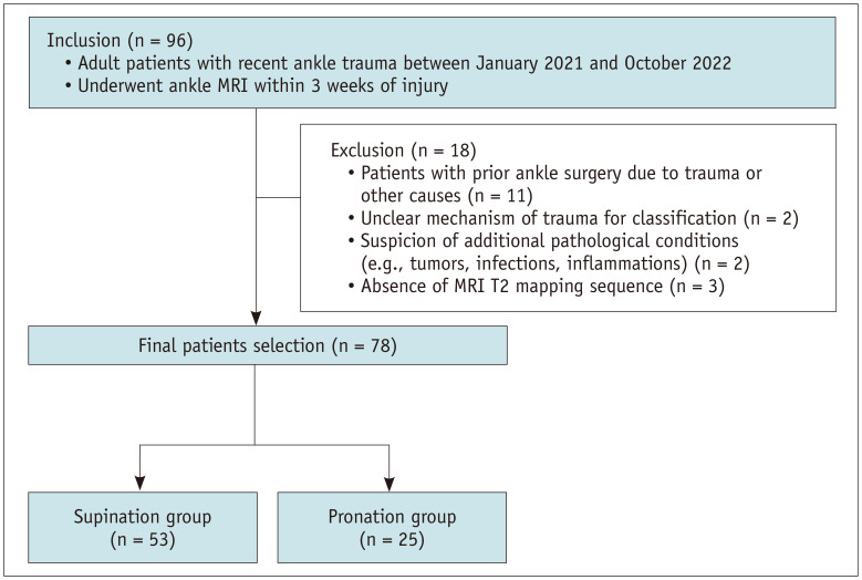
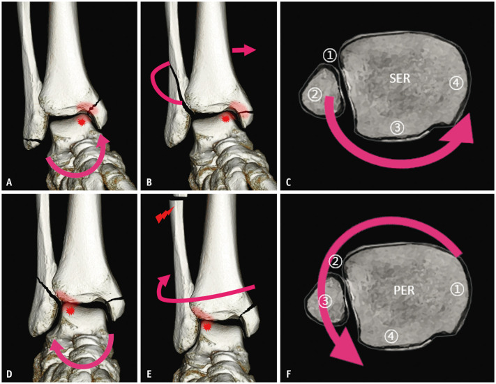
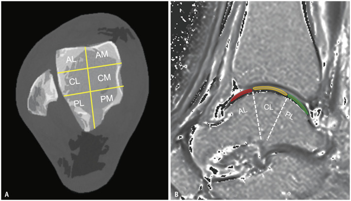
Materials and methods: This retrospective study analyzed 78 patients who underwent ankle MRI with T2 mapping for acute ankle trauma between January 2021 and October 2022. Patients were classified into the supination (S) and pronation (P) groups based on the LH classification, and then divided into subgroups based on posterior malleolus (PM) involvement. The T2 values for the talar cartilage were quantitatively measured in six anatomical regions defined by the combination of medial vs. lateral and anterior vs. central vs. posterior. The T2 mapping values in each region of the talus were compared between the S and P groups and between the PM and non-PM injury groups using t-tests. The T2 values were also compared between the medial and lateral sides within each group.
Results: Among the 78 patients (mean age, 38.62 ± 14.82 years; 47 male), 53 and 25 were in the S and P groups, respectively, and 53 patients showed PM involvement. In comparison with the P group, the S group exhibited higher T2 values in the medial portion (61.27 ± 8.30 vs. 54.03 ± 6.96; P < 0.001) and lower T2 values in the lateral talus (54.95 ± 8.47 vs. 64.15 ± 7.31; P < 0.001). The PM injury group showed higher T2 values in the posterior region than the non-PM injury group (P ≤ 0.011). Within the PM injury group, T2 values were higher in the anteromedial and posterolateral regions than on the opposite sides (P = 0.037 and 0.011, respectively).
Conclusion: MRI T2 values demonstrated significant regional variations in the talar dome cartilage in acute ankle trauma, and the T2 values may reflect different ankle trauma mechanisms and PM involvement. Thus, T2 mapping can facilitate evaluation of talar cartilage alterations.
期刊介绍:
The inaugural issue of the Korean J Radiol came out in March 2000. Our journal aims to produce and propagate knowledge on radiologic imaging and related sciences.
A unique feature of the articles published in the Journal will be their reflection of global trends in radiology combined with an East-Asian perspective. Geographic differences in disease prevalence will be reflected in the contents of papers, and this will serve to enrich our body of knowledge.
World''s outstanding radiologists from many countries are serving as editorial board of our journal.

 求助内容:
求助内容: 应助结果提醒方式:


