{"title":"Repeated Gonadotropin Administration Suppresses T Cell Development in the Mouse Thymus.","authors":"Jin Yoon, Sojung Sun, Soeun Moon, Hyunwon Yang","doi":"10.12717/DR.2025.29.1.1","DOIUrl":null,"url":null,"abstract":"<p><p>Gonadotropins, such as follicle-stimulating hormone (FSH) and human chorionic gonadotropin (hCG), are widely used to induce ovarian hyperovulation during in vitro fertilization and embryo transfer (IVF-ET) for the treatment of infertility. However, the effects of repeated administration of these gonadotropins on immune function, particularly on T cell development in the thymus, remain poorly understood. This study investigated the effects of repeated administration of pregnant mare serum gonadotropin (PMSG) and hCG on thymic T cell development in mice. Histological analysis revealed structural changes in the thymus, including a blurred boundary between the medulla and cortex and reduced vascularization after repeated administration of PMSG and hCG. Quantitative real-time PCR showed increased expression of adipogenesis-related genes [phosphoenolpyruvate carboxykinase (PEPCK), adipocyte fatty acid-binding protein 2 (aP2), peroxisome proliferator-activated receptor gamma (PPARγ)] but no significant changes in thymic epithelial cell-related genes [autoimmune regulator (AIRE), epithelial V-like antigen (EVA), interleukin 7 (IL-7)]. Flow cytometry revealed a decrease in CD4<sup>+</sup>CD8<sup>+</sup> T cells and an increase in CD4-CD8-T cells with altered CD25/CD44 subsets. In addition, CD4<sup>+</sup> and CD8<sup>+</sup> T cells in the spleen were significantly reduced. These findings suggest that repeated gonadotropin exposure may disrupt thymic T cell development and peripheral T cell populations, potentially impairing immune function. Further research is needed to elucidate the underlying mechanisms and broader immunologic consequences of gonadotropin use in infertility treatment.</p>","PeriodicalId":72791,"journal":{"name":"Development & reproduction","volume":"29 1","pages":"1-11"},"PeriodicalIF":0.0000,"publicationDate":"2025-03-01","publicationTypes":"Journal Article","fieldsOfStudy":null,"isOpenAccess":false,"openAccessPdf":"https://www.ncbi.nlm.nih.gov/pmc/articles/PMC12004010/pdf/","citationCount":"0","resultStr":null,"platform":"Semanticscholar","paperid":null,"PeriodicalName":"Development & reproduction","FirstCategoryId":"1085","ListUrlMain":"https://doi.org/10.12717/DR.2025.29.1.1","RegionNum":0,"RegionCategory":null,"ArticlePicture":[],"TitleCN":null,"AbstractTextCN":null,"PMCID":null,"EPubDate":"2025/3/31 0:00:00","PubModel":"Epub","JCR":"","JCRName":"","Score":null,"Total":0}
引用次数: 0
Abstract
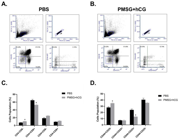
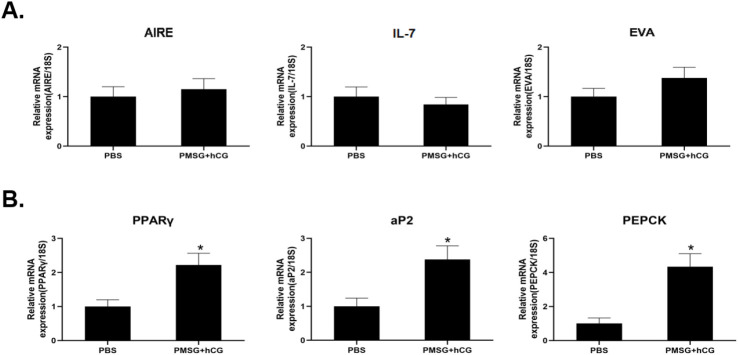
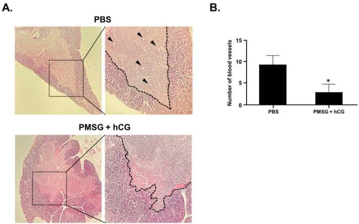
Gonadotropins, such as follicle-stimulating hormone (FSH) and human chorionic gonadotropin (hCG), are widely used to induce ovarian hyperovulation during in vitro fertilization and embryo transfer (IVF-ET) for the treatment of infertility. However, the effects of repeated administration of these gonadotropins on immune function, particularly on T cell development in the thymus, remain poorly understood. This study investigated the effects of repeated administration of pregnant mare serum gonadotropin (PMSG) and hCG on thymic T cell development in mice. Histological analysis revealed structural changes in the thymus, including a blurred boundary between the medulla and cortex and reduced vascularization after repeated administration of PMSG and hCG. Quantitative real-time PCR showed increased expression of adipogenesis-related genes [phosphoenolpyruvate carboxykinase (PEPCK), adipocyte fatty acid-binding protein 2 (aP2), peroxisome proliferator-activated receptor gamma (PPARγ)] but no significant changes in thymic epithelial cell-related genes [autoimmune regulator (AIRE), epithelial V-like antigen (EVA), interleukin 7 (IL-7)]. Flow cytometry revealed a decrease in CD4+CD8+ T cells and an increase in CD4-CD8-T cells with altered CD25/CD44 subsets. In addition, CD4+ and CD8+ T cells in the spleen were significantly reduced. These findings suggest that repeated gonadotropin exposure may disrupt thymic T cell development and peripheral T cell populations, potentially impairing immune function. Further research is needed to elucidate the underlying mechanisms and broader immunologic consequences of gonadotropin use in infertility treatment.

 求助内容:
求助内容: 应助结果提醒方式:


