Othman Zuhir, Muzaffar Apipi, Jaswinder Singh, Yee Chin Lim
{"title":"Suspected Emphysematous Cellulitis of the Face Secondary to Untreated Nondisplaced Angle of the Mandible Fracture: A Case Report.","authors":"Othman Zuhir, Muzaffar Apipi, Jaswinder Singh, Yee Chin Lim","doi":"10.1155/crid/6247721","DOIUrl":null,"url":null,"abstract":"<p><p>Facial cellulitis with palpable crepitus is a rare complication following a nondisplaced angle of mandible fracture. We report a case of a male in his mid-20s who presented with suspected emphysematous cellulitis of the face 3 days after an assault. Clinical examination revealed diffuse facial swelling with palpable subcutaneous crepitus over the left cheek and submandibular region. The patient had trismus and suppuration of the lower left third molar but no intraoral soft tissue injury. An orthopantomogram (OPG) showed a left nondisplaced angle of mandible fracture and multilocular bubble-like radiolucencies, suggesting submasseteric and submandibular gas accumulation. Due to financial constraints, advanced imaging and histological evaluation were not performed, limiting diagnostic certainty. Incision and drainage were performed, followed by intermaxillary fixation (IMF), and the infection was resolved with intravenous antibiotics. Open reduction internal fixation (ORIF) was not pursued due to cost limitations, but the fracture healed successfully within 6 weeks of IMF. This case underscores the importance of recognising gas-forming infections following mandibular trauma, the diagnostic challenges in resource-limited settings, and the role of early intervention in preventing severe complications.</p>","PeriodicalId":46841,"journal":{"name":"Case Reports in Dentistry","volume":"2025 ","pages":"6247721"},"PeriodicalIF":0.9000,"publicationDate":"2025-04-21","publicationTypes":"Journal Article","fieldsOfStudy":null,"isOpenAccess":false,"openAccessPdf":"https://www.ncbi.nlm.nih.gov/pmc/articles/PMC12037252/pdf/","citationCount":"0","resultStr":null,"platform":"Semanticscholar","paperid":null,"PeriodicalName":"Case Reports in Dentistry","FirstCategoryId":"1085","ListUrlMain":"https://doi.org/10.1155/crid/6247721","RegionNum":0,"RegionCategory":null,"ArticlePicture":[],"TitleCN":null,"AbstractTextCN":null,"PMCID":null,"EPubDate":"2025/1/1 0:00:00","PubModel":"eCollection","JCR":"Q4","JCRName":"DENTISTRY, ORAL SURGERY & MEDICINE","Score":null,"Total":0}
引用次数: 0
Abstract
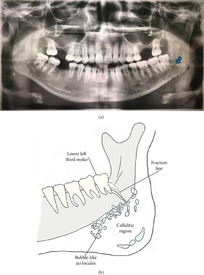
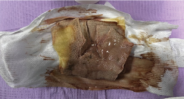
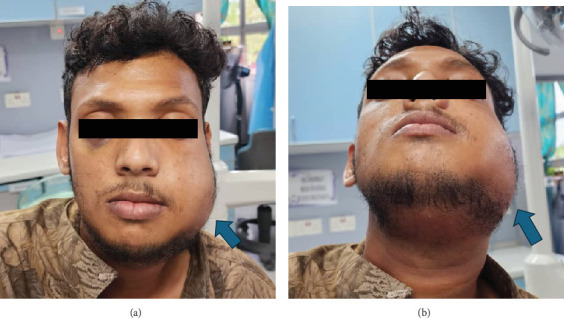
Facial cellulitis with palpable crepitus is a rare complication following a nondisplaced angle of mandible fracture. We report a case of a male in his mid-20s who presented with suspected emphysematous cellulitis of the face 3 days after an assault. Clinical examination revealed diffuse facial swelling with palpable subcutaneous crepitus over the left cheek and submandibular region. The patient had trismus and suppuration of the lower left third molar but no intraoral soft tissue injury. An orthopantomogram (OPG) showed a left nondisplaced angle of mandible fracture and multilocular bubble-like radiolucencies, suggesting submasseteric and submandibular gas accumulation. Due to financial constraints, advanced imaging and histological evaluation were not performed, limiting diagnostic certainty. Incision and drainage were performed, followed by intermaxillary fixation (IMF), and the infection was resolved with intravenous antibiotics. Open reduction internal fixation (ORIF) was not pursued due to cost limitations, but the fracture healed successfully within 6 weeks of IMF. This case underscores the importance of recognising gas-forming infections following mandibular trauma, the diagnostic challenges in resource-limited settings, and the role of early intervention in preventing severe complications.
期刊介绍:
Case Reports in Dentistry is a peer-reviewed, Open Access journal that publishes case reports and case series in all areas of dentistry, including periodontal diseases, dental implants, oral pathology, as well as oral and maxillofacial surgery.

 求助内容:
求助内容: 应助结果提醒方式:


