{"title":"A case of phosphoglyceride crystal deposition disease in the maxilla.","authors":"Emi Saitou, Takaaki Sano, Mai Seki, Takahiro Yamaguchi, Tetsunari Oyama, Satoshi Yokoo","doi":"10.1186/s13000-025-01641-2","DOIUrl":null,"url":null,"abstract":"<p><strong>Background: </strong>Phosphoglyceride crystal deposition disease (PCDD) is a rare disorder in which phosphoglyceride crystals accumulate in soft tissues and bones. It tends to occur years after surgery, trauma, or repeated injections.</p><p><strong>Case presentation: </strong>An 81-year-old woman was referred to our department because of swelling of the left maxillary gingiva. The left maxillary second molar had been extracted more than 10 years earlier. Surgical biopsy was performed, and histopathological findings indicated a foreign body granuloma. The patient underwent tumorectomy, during which we found a yellowish tumor. The pathologic findings were the characteristic crystal deposition, fibril-like crystals, and giant cells around the crystals. Gold hydroxamic acid staining revealed positivity for the crystals. The final pathological diagnosis was PCDD. The patient had no further symptoms and no disease recurrence.</p><p><strong>Conclusions: </strong>It is relatively easy to diagnose PCDD from the characteristic histopathological findings; however, it may be overlooked by pathologists who are unaware of the disease. T2-weighted magnetic resonance imaging of PCDD in the jawbone has depicted low intensity, a finding that differs from those of ordinary cancers and odontogenic tumors. The oral cavity often undergoes surgical procedures, and PCDD may form, and grow.</p>","PeriodicalId":11237,"journal":{"name":"Diagnostic Pathology","volume":"20 1","pages":"45"},"PeriodicalIF":2.3000,"publicationDate":"2025-04-16","publicationTypes":"Journal Article","fieldsOfStudy":null,"isOpenAccess":false,"openAccessPdf":"https://www.ncbi.nlm.nih.gov/pmc/articles/PMC12004582/pdf/","citationCount":"0","resultStr":null,"platform":"Semanticscholar","paperid":null,"PeriodicalName":"Diagnostic Pathology","FirstCategoryId":"3","ListUrlMain":"https://doi.org/10.1186/s13000-025-01641-2","RegionNum":3,"RegionCategory":"医学","ArticlePicture":[],"TitleCN":null,"AbstractTextCN":null,"PMCID":null,"EPubDate":"","PubModel":"","JCR":"Q2","JCRName":"PATHOLOGY","Score":null,"Total":0}
引用次数: 0
Abstract
Background: Phosphoglyceride crystal deposition disease (PCDD) is a rare disorder in which phosphoglyceride crystals accumulate in soft tissues and bones. It tends to occur years after surgery, trauma, or repeated injections.
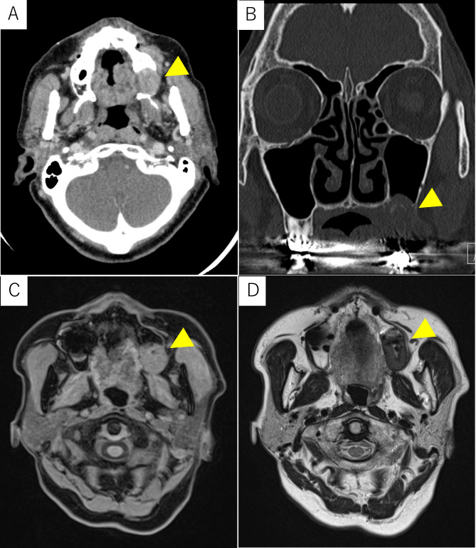
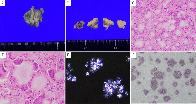
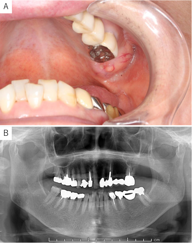
Case presentation: An 81-year-old woman was referred to our department because of swelling of the left maxillary gingiva. The left maxillary second molar had been extracted more than 10 years earlier. Surgical biopsy was performed, and histopathological findings indicated a foreign body granuloma. The patient underwent tumorectomy, during which we found a yellowish tumor. The pathologic findings were the characteristic crystal deposition, fibril-like crystals, and giant cells around the crystals. Gold hydroxamic acid staining revealed positivity for the crystals. The final pathological diagnosis was PCDD. The patient had no further symptoms and no disease recurrence.
Conclusions: It is relatively easy to diagnose PCDD from the characteristic histopathological findings; however, it may be overlooked by pathologists who are unaware of the disease. T2-weighted magnetic resonance imaging of PCDD in the jawbone has depicted low intensity, a finding that differs from those of ordinary cancers and odontogenic tumors. The oral cavity often undergoes surgical procedures, and PCDD may form, and grow.
期刊介绍:
Diagnostic Pathology is an open access, peer-reviewed, online journal that considers research in surgical and clinical pathology, immunology, and biology, with a special focus on cutting-edge approaches in diagnostic pathology and tissue-based therapy. The journal covers all aspects of surgical pathology, including classic diagnostic pathology, prognosis-related diagnosis (tumor stages, prognosis markers, such as MIB-percentage, hormone receptors, etc.), and therapy-related findings. The journal also focuses on the technological aspects of pathology, including molecular biology techniques, morphometry aspects (stereology, DNA analysis, syntactic structure analysis), communication aspects (telecommunication, virtual microscopy, virtual pathology institutions, etc.), and electronic education and quality assurance (for example interactive publication, on-line references with automated updating, etc.).

 求助内容:
求助内容: 应助结果提醒方式:


