{"title":"Exploring the varied expressions of basic fibroblast growth factor in different histopathological grades of oral submucous fibrosis.","authors":"Geetpriya Kaur, Vijay Wadhwan, Kiran Kumar, Aparna Pathak, Farnaz Y Shah, Pallak Arora","doi":"10.4103/jomfp.jomfp_173_24","DOIUrl":null,"url":null,"abstract":"<p><strong>Context: </strong>Oral submucous fibrosis (OSMF) is a chronic disorder with multi-factorial aetiology. The OSMF pathophysiology includes the homeostatic equilibrium disruption between synthesis and degradation of extracellular matrix. Thus, various growth factors produced by activated inflammatory cells may promote fibrosis by inducing fibroblast proliferation, collagen synthesis upregulation, and reduced collagenase production.</p><p><strong>Aims: </strong>To correlate the role of basic fibroblast growth factor (bFGF) in the endothelial cells, fibroblasts, and connective tissue stroma in varying grades of OSMF. The bFGF expression was also correlated with the amount of inflammation.</p><p><strong>Settings and design: </strong>This retrospective study was designed to evaluate bFGF expression in 30 histopathologically diagnosed cases of OSMF from the Department of Oral and Maxillofacial Pathology, I.T.S CDSR, Muradnagar.</p><p><strong>Materials and methods: </strong>We included 30 cases, ten each of early, intermediate, and advanced stages of OSMF. Immunohistochemical staining using bFGF antibody was performed, and bFGF expression was noted in the blood vessels, fibroblasts, and connective tissue stroma in all the study cases.</p><p><strong>Statistical analysis used: </strong>Different variables were analysed using the ANOVA test, <i>post</i> <i>hoc</i> test, and Bonferroni test.</p><p><strong>Results: </strong>The bFGF-labelled blood vessels and fibroblasts were significantly higher in early OSMF cases than in the intermediate and advanced groups. bFGF expression was significantly observed in the connective tissue stroma in most of the cases.</p><p><strong>Conclusions: </strong>The bFGF intensity was mild, moderate, and severe in early, intermediate, and advanced OSMF cases, respectively. Moreover, bFGF expression was noted in the blood vessels, fibroblasts, and connective tissue stroma in the majority of the cases.</p>","PeriodicalId":38846,"journal":{"name":"Journal of Oral and Maxillofacial Pathology","volume":"29 1","pages":"76-80"},"PeriodicalIF":0.0000,"publicationDate":"2025-01-01","publicationTypes":"Journal Article","fieldsOfStudy":null,"isOpenAccess":false,"openAccessPdf":"https://www.ncbi.nlm.nih.gov/pmc/articles/PMC12002573/pdf/","citationCount":"0","resultStr":null,"platform":"Semanticscholar","paperid":null,"PeriodicalName":"Journal of Oral and Maxillofacial Pathology","FirstCategoryId":"1085","ListUrlMain":"https://doi.org/10.4103/jomfp.jomfp_173_24","RegionNum":0,"RegionCategory":null,"ArticlePicture":[],"TitleCN":null,"AbstractTextCN":null,"PMCID":null,"EPubDate":"2025/3/28 0:00:00","PubModel":"Epub","JCR":"Q3","JCRName":"Medicine","Score":null,"Total":0}
引用次数: 0
Abstract
Context: Oral submucous fibrosis (OSMF) is a chronic disorder with multi-factorial aetiology. The OSMF pathophysiology includes the homeostatic equilibrium disruption between synthesis and degradation of extracellular matrix. Thus, various growth factors produced by activated inflammatory cells may promote fibrosis by inducing fibroblast proliferation, collagen synthesis upregulation, and reduced collagenase production.
Aims: To correlate the role of basic fibroblast growth factor (bFGF) in the endothelial cells, fibroblasts, and connective tissue stroma in varying grades of OSMF. The bFGF expression was also correlated with the amount of inflammation.
Settings and design: This retrospective study was designed to evaluate bFGF expression in 30 histopathologically diagnosed cases of OSMF from the Department of Oral and Maxillofacial Pathology, I.T.S CDSR, Muradnagar.
Materials and methods: We included 30 cases, ten each of early, intermediate, and advanced stages of OSMF. Immunohistochemical staining using bFGF antibody was performed, and bFGF expression was noted in the blood vessels, fibroblasts, and connective tissue stroma in all the study cases.
Statistical analysis used: Different variables were analysed using the ANOVA test, posthoc test, and Bonferroni test.
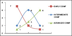
Results: The bFGF-labelled blood vessels and fibroblasts were significantly higher in early OSMF cases than in the intermediate and advanced groups. bFGF expression was significantly observed in the connective tissue stroma in most of the cases.
Conclusions: The bFGF intensity was mild, moderate, and severe in early, intermediate, and advanced OSMF cases, respectively. Moreover, bFGF expression was noted in the blood vessels, fibroblasts, and connective tissue stroma in the majority of the cases.
期刊介绍:
The journal of Oral and Maxillofacial Pathology [ISSN:print-(0973-029X, online-1998-393X)] is a tri-annual journal published on behalf of “The Indian Association of Oral and Maxillofacial Pathologists” (IAOMP). The publication of JOMFP was started in the year 1993. The journal publishes papers on a wide spectrum of topics associated with the scope of Oral and Maxillofacial Pathology, also, ensuring scientific merit and quality. It is a comprehensive reading material for the professionals who want to upgrade their diagnostic skills in Oral Diseases; allows exposure to newer topics and methods of research in the Oral-facial Tissues and Pathology. New features allow an open minded thinking and approach to various pathologies. It also encourages authors to showcase quality work done by them and to compile relevant cases which are diagnostically challenging. The Journal takes pride in maintaining the quality of articles and photomicrographs.

 求助内容:
求助内容: 应助结果提醒方式:


