{"title":"Increased CT Attenuation of Pericolic Adipose Tissue as a Noninvasive Marker of Disease Severity in Ulcerative Colitis.","authors":"Jun Lu, Hui Xu, Jing Zheng, Tianxin Cheng, Xinjun Han, Yuxin Wang, Xuxu Meng, Xiaoyang Li, Jiahui Jiang, Xue Dong, Xijie Zhang, Zhenchang Wang, Zhenghan Yang, Lixue Xu","doi":"10.3348/kjr.2024.0857","DOIUrl":null,"url":null,"abstract":"<p><strong>Objective: </strong>Accurate evaluation of inflammation severity in ulcerative colitis (UC) can guide treatment strategy selection. The potential value of the pericolic fat attenuation index (FAI) on CT as an indicator of disease severity remains unknown. This study aimed to assess the diagnostic accuracy of pericolic FAI in predicting UC severity.</p><p><strong>Materials and methods: </strong>This retrospective study enrolled 148 patients (mean age 48 years; 87 males). The fat attenuation on CT was measured in four different locations: the mesocolic vascular side (MS) and opposite side of MS (OMS) around the most severe bowel lesion, the retroperitoneal space (RS), and the subcutaneous area. The fat attenuation indices (FAI<sub>MS</sub>, FAI<sub>OMS</sub>, and FAI<sub>RS</sub>) were calculated as the fat attenuation measured in MS, OMS, and RS, respectively, minus that of the subcutaneous area, and were obtained in the non-enhanced, arterial, and delayed phases. Correlations between the FAI and UC Endoscopic Index of Severity (UCEIS) were assessed using Spearman's correlation. Predictors of severe UC (UCEIS ≥7) were selected by univariable analysis. The performance of FAI in predicting severe UC was evaluated using the area under the receiver operating characteristic curve (AUC).</p><p><strong>Results: </strong>The FAI<sub>MS</sub> and FAI<sub>OMS</sub> scores were significantly higher than FAI<sub>RS</sub> in three phases (all <i>P</i> < 0.001). The FAI<sub>MS</sub> and FAI<sub>OMS</sub> scores moderately correlated with the UCEIS score (<i>r</i> = 0.474-0.649 among the three phases). Additionally, FAI<sub>MS</sub> and FAI<sub>OMS</sub> identified severe UC, with AUC varying from 0.77 to 0.85.</p><p><strong>Conclusion: </strong>Increased CT attenuation of pericolic adipose tissue could serve as a noninvasive marker for evaluating UC severity. FAI<sub>MS</sub> and FAI<sub>OMS</sub> of three phases showed similar prediction accuracies for severe UC identification.</p>","PeriodicalId":17881,"journal":{"name":"Korean Journal of Radiology","volume":"26 5","pages":"411-421"},"PeriodicalIF":5.3000,"publicationDate":"2025-05-01","publicationTypes":"Journal Article","fieldsOfStudy":null,"isOpenAccess":false,"openAccessPdf":"https://www.ncbi.nlm.nih.gov/pmc/articles/PMC12055264/pdf/","citationCount":"0","resultStr":null,"platform":"Semanticscholar","paperid":null,"PeriodicalName":"Korean Journal of Radiology","FirstCategoryId":"3","ListUrlMain":"https://doi.org/10.3348/kjr.2024.0857","RegionNum":2,"RegionCategory":"医学","ArticlePicture":[],"TitleCN":null,"AbstractTextCN":null,"PMCID":null,"EPubDate":"","PubModel":"","JCR":"Q1","JCRName":"RADIOLOGY, NUCLEAR MEDICINE & MEDICAL IMAGING","Score":null,"Total":0}
引用次数: 0
Abstract
Objective: Accurate evaluation of inflammation severity in ulcerative colitis (UC) can guide treatment strategy selection. The potential value of the pericolic fat attenuation index (FAI) on CT as an indicator of disease severity remains unknown. This study aimed to assess the diagnostic accuracy of pericolic FAI in predicting UC severity.
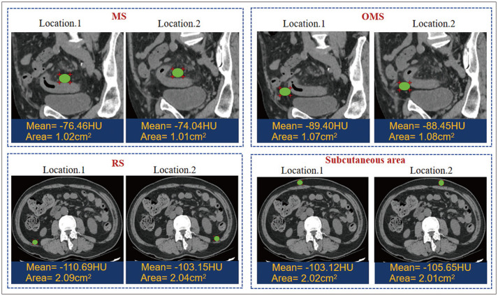
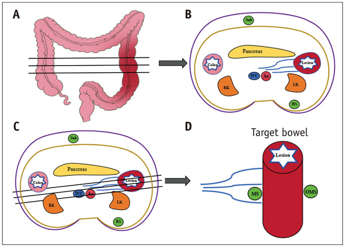
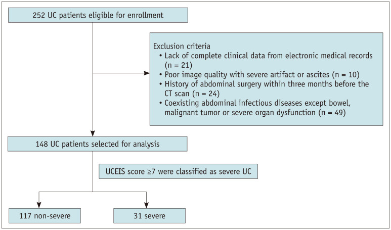
Materials and methods: This retrospective study enrolled 148 patients (mean age 48 years; 87 males). The fat attenuation on CT was measured in four different locations: the mesocolic vascular side (MS) and opposite side of MS (OMS) around the most severe bowel lesion, the retroperitoneal space (RS), and the subcutaneous area. The fat attenuation indices (FAIMS, FAIOMS, and FAIRS) were calculated as the fat attenuation measured in MS, OMS, and RS, respectively, minus that of the subcutaneous area, and were obtained in the non-enhanced, arterial, and delayed phases. Correlations between the FAI and UC Endoscopic Index of Severity (UCEIS) were assessed using Spearman's correlation. Predictors of severe UC (UCEIS ≥7) were selected by univariable analysis. The performance of FAI in predicting severe UC was evaluated using the area under the receiver operating characteristic curve (AUC).
Results: The FAIMS and FAIOMS scores were significantly higher than FAIRS in three phases (all P < 0.001). The FAIMS and FAIOMS scores moderately correlated with the UCEIS score (r = 0.474-0.649 among the three phases). Additionally, FAIMS and FAIOMS identified severe UC, with AUC varying from 0.77 to 0.85.
Conclusion: Increased CT attenuation of pericolic adipose tissue could serve as a noninvasive marker for evaluating UC severity. FAIMS and FAIOMS of three phases showed similar prediction accuracies for severe UC identification.
期刊介绍:
The inaugural issue of the Korean J Radiol came out in March 2000. Our journal aims to produce and propagate knowledge on radiologic imaging and related sciences.
A unique feature of the articles published in the Journal will be their reflection of global trends in radiology combined with an East-Asian perspective. Geographic differences in disease prevalence will be reflected in the contents of papers, and this will serve to enrich our body of knowledge.
World''s outstanding radiologists from many countries are serving as editorial board of our journal.

 求助内容:
求助内容: 应助结果提醒方式:


