Christian T Schamberger, Arnold J Suda, Tobias Grossner, Gerhard Schmidmaier, Stephan Stein
{"title":"Sonography-based determination of hip joint anterior head-neck offset is reliable and reproducible for CAM deformity assessment.","authors":"Christian T Schamberger, Arnold J Suda, Tobias Grossner, Gerhard Schmidmaier, Stephan Stein","doi":"10.1055/a-2537-7181","DOIUrl":null,"url":null,"abstract":"<p><strong>Purpose: </strong>Native X-ray, magnetic resonance imaging (MRI), and computed tomography (CT) are standard methods for determining head-neck offset (HNO) in femoro-acetabular impingement (FAI). Our hypothesis was that sonography-assisted determination of the offset in CAM deformity of the hip is a cheap, radiation-free, and reliable alternative to conventional alpha-angle determination.</p><p><strong>Methods: </strong>Patients with hip pain and suspected CAM impingement who underwent anterior-longitudinal hip sonography according to DEGUM standard procedures and MRI were included in this single-center study between January 2015 and December 2019. Offset was determined three times on MRI and sonography by two independent investigators.</p><p><strong>Results: </strong>285 patients were screened and 110 patients (49 females, 61 males) met the inclusion criteria. The mean age at the time of investigation of 54 left and 56 right hip joints was 54.2 years. 1320 measurements were performed. No significant difference in HNO determination between MRI (6.11 mm+/-2.37) and sonography (5.93 mm+/-2.20) could be identified. The mean difference was 0.32 mm+/-0.32 mm (p>0.05) with a maximum deviation of 2.08 mm (outlier).</p><p><strong>Conclusion: </strong>Sonography-assisted determination of head-neck offset is a reliable and reproducible method and is not inferior to determination with MRI. Sonography can be used initially as an alternative or additional tool for the qualitative determination of CAM deformity of the hip joint.</p>","PeriodicalId":44852,"journal":{"name":"Ultrasound International Open","volume":"11 ","pages":"a25377181"},"PeriodicalIF":1.6000,"publicationDate":"2025-04-24","publicationTypes":"Journal Article","fieldsOfStudy":null,"isOpenAccess":false,"openAccessPdf":"https://www.ncbi.nlm.nih.gov/pmc/articles/PMC12039884/pdf/","citationCount":"0","resultStr":null,"platform":"Semanticscholar","paperid":null,"PeriodicalName":"Ultrasound International Open","FirstCategoryId":"1085","ListUrlMain":"https://doi.org/10.1055/a-2537-7181","RegionNum":0,"RegionCategory":null,"ArticlePicture":[],"TitleCN":null,"AbstractTextCN":null,"PMCID":null,"EPubDate":"2025/1/1 0:00:00","PubModel":"eCollection","JCR":"Q3","JCRName":"RADIOLOGY, NUCLEAR MEDICINE & MEDICAL IMAGING","Score":null,"Total":0}
引用次数: 0
Abstract
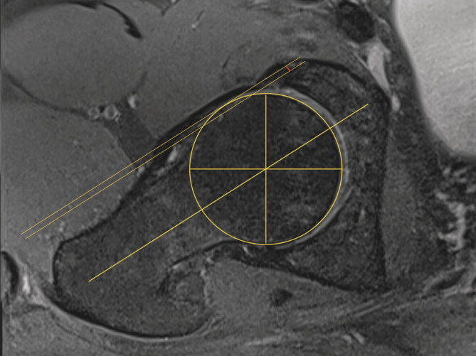
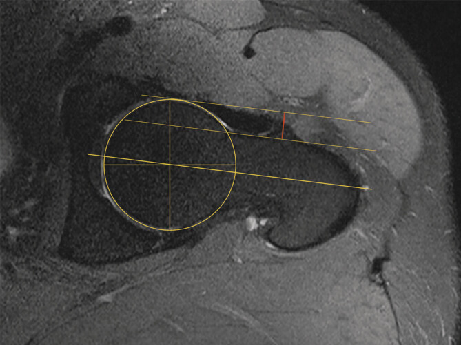
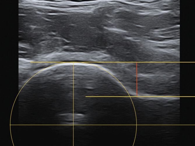
Purpose: Native X-ray, magnetic resonance imaging (MRI), and computed tomography (CT) are standard methods for determining head-neck offset (HNO) in femoro-acetabular impingement (FAI). Our hypothesis was that sonography-assisted determination of the offset in CAM deformity of the hip is a cheap, radiation-free, and reliable alternative to conventional alpha-angle determination.
Methods: Patients with hip pain and suspected CAM impingement who underwent anterior-longitudinal hip sonography according to DEGUM standard procedures and MRI were included in this single-center study between January 2015 and December 2019. Offset was determined three times on MRI and sonography by two independent investigators.
Results: 285 patients were screened and 110 patients (49 females, 61 males) met the inclusion criteria. The mean age at the time of investigation of 54 left and 56 right hip joints was 54.2 years. 1320 measurements were performed. No significant difference in HNO determination between MRI (6.11 mm+/-2.37) and sonography (5.93 mm+/-2.20) could be identified. The mean difference was 0.32 mm+/-0.32 mm (p>0.05) with a maximum deviation of 2.08 mm (outlier).
Conclusion: Sonography-assisted determination of head-neck offset is a reliable and reproducible method and is not inferior to determination with MRI. Sonography can be used initially as an alternative or additional tool for the qualitative determination of CAM deformity of the hip joint.

 求助内容:
求助内容: 应助结果提醒方式:


