Basheer Abdullah Marzoog, Peter Chomakhidze, Daria Gognieva, Artemiy Silantyev, Alexander Suvorov, Magomed Abdullaev, Natalia Mozzhukhina, Darya Alexandrovna Filippova, Sergey Vladimirovich Kostin, Maria Kolpashnikova, Natalya Ershova, Nikolay Ushakov, Dinara Mesitskaya, Philipp Kopylov
{"title":"Development and validation of a machine learning model for diagnosis of ischemic heart disease using single-lead electrocardiogram parameters.","authors":"Basheer Abdullah Marzoog, Peter Chomakhidze, Daria Gognieva, Artemiy Silantyev, Alexander Suvorov, Magomed Abdullaev, Natalia Mozzhukhina, Darya Alexandrovna Filippova, Sergey Vladimirovich Kostin, Maria Kolpashnikova, Natalya Ershova, Nikolay Ushakov, Dinara Mesitskaya, Philipp Kopylov","doi":"10.4330/wjc.v17.i4.104396","DOIUrl":null,"url":null,"abstract":"<p><strong>Background: </strong>Ischemic heart disease (IHD) impacts the quality of life and has the highest mortality rate of cardiovascular diseases globally.</p><p><strong>Aim: </strong>To compare variations in the parameters of the single-lead electrocardiogram (ECG) during resting conditions and physical exertion in individuals diagnosed with IHD and those without the condition using vasodilator-induced stress computed tomography (CT) myocardial perfusion imaging as the diagnostic reference standard.</p><p><strong>Methods: </strong>This single center observational study included 80 participants. The participants were aged ≥ 40 years and given an informed written consent to participate in the study. Both groups, G1 (<i>n</i> = 31) with and G2 (<i>n</i> = 49) without post stress induced myocardial perfusion defect, passed cardiologist consultation, anthropometric measurements, blood pressure and pulse rate measurement, echocardiography, cardio-ankle vascular index, bicycle ergometry, recording 3-min single-lead ECG (Cardio-Qvark) before and just after bicycle ergometry followed by performing CT myocardial perfusion. The LASSO regression with nested cross-validation was used to find the association between Cardio-Qvark parameters and the existence of the perfusion defect. Statistical processing was performed with the R programming language v4.2, Python v.3.10 [^R], and Statistica 12 program.</p><p><strong>Results: </strong>Bicycle ergometry yielded an area under the receiver operating characteristic curve of 50.7% [95% confidence interval (CI): 0.388-0.625], specificity of 53.1% (95%CI: 0.392-0.673), and sensitivity of 48.4% (95%CI: 0.306-0.657). In contrast, the Cardio-Qvark test performed notably better with an area under the receiver operating characteristic curve of 67% (95%CI: 0.530-0.801), specificity of 75.5% (95%CI: 0.628-0.88), and sensitivity of 51.6% (95%CI: 0.333-0.695).</p><p><strong>Conclusion: </strong>The single-lead ECG has a relatively higher diagnostic accuracy compared with bicycle ergometry by using machine learning models, but the difference was not statistically significant. However, further investigations are required to uncover the hidden capabilities of single-lead ECG in IHD diagnosis.</p>","PeriodicalId":23800,"journal":{"name":"World Journal of Cardiology","volume":"17 4","pages":"104396"},"PeriodicalIF":2.8000,"publicationDate":"2025-04-26","publicationTypes":"Journal Article","fieldsOfStudy":null,"isOpenAccess":false,"openAccessPdf":"https://www.ncbi.nlm.nih.gov/pmc/articles/PMC12038698/pdf/","citationCount":"0","resultStr":null,"platform":"Semanticscholar","paperid":null,"PeriodicalName":"World Journal of Cardiology","FirstCategoryId":"1085","ListUrlMain":"https://doi.org/10.4330/wjc.v17.i4.104396","RegionNum":0,"RegionCategory":null,"ArticlePicture":[],"TitleCN":null,"AbstractTextCN":null,"PMCID":null,"EPubDate":"","PubModel":"","JCR":"Q3","JCRName":"CARDIAC & CARDIOVASCULAR SYSTEMS","Score":null,"Total":0}
引用次数: 0
Abstract
Background: Ischemic heart disease (IHD) impacts the quality of life and has the highest mortality rate of cardiovascular diseases globally.
Aim: To compare variations in the parameters of the single-lead electrocardiogram (ECG) during resting conditions and physical exertion in individuals diagnosed with IHD and those without the condition using vasodilator-induced stress computed tomography (CT) myocardial perfusion imaging as the diagnostic reference standard.
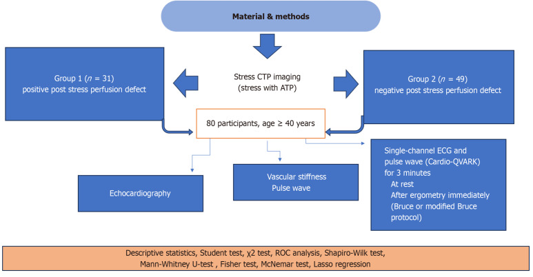
Methods: This single center observational study included 80 participants. The participants were aged ≥ 40 years and given an informed written consent to participate in the study. Both groups, G1 (n = 31) with and G2 (n = 49) without post stress induced myocardial perfusion defect, passed cardiologist consultation, anthropometric measurements, blood pressure and pulse rate measurement, echocardiography, cardio-ankle vascular index, bicycle ergometry, recording 3-min single-lead ECG (Cardio-Qvark) before and just after bicycle ergometry followed by performing CT myocardial perfusion. The LASSO regression with nested cross-validation was used to find the association between Cardio-Qvark parameters and the existence of the perfusion defect. Statistical processing was performed with the R programming language v4.2, Python v.3.10 [^R], and Statistica 12 program.
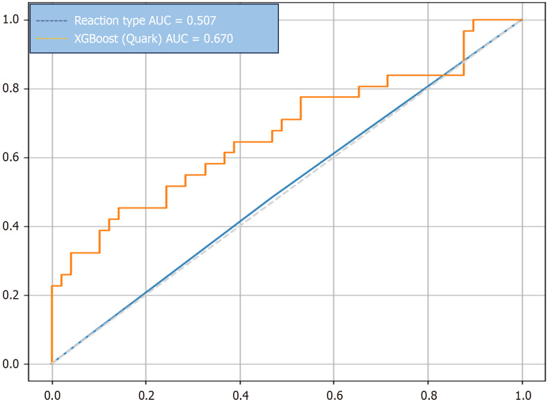
Results: Bicycle ergometry yielded an area under the receiver operating characteristic curve of 50.7% [95% confidence interval (CI): 0.388-0.625], specificity of 53.1% (95%CI: 0.392-0.673), and sensitivity of 48.4% (95%CI: 0.306-0.657). In contrast, the Cardio-Qvark test performed notably better with an area under the receiver operating characteristic curve of 67% (95%CI: 0.530-0.801), specificity of 75.5% (95%CI: 0.628-0.88), and sensitivity of 51.6% (95%CI: 0.333-0.695).
Conclusion: The single-lead ECG has a relatively higher diagnostic accuracy compared with bicycle ergometry by using machine learning models, but the difference was not statistically significant. However, further investigations are required to uncover the hidden capabilities of single-lead ECG in IHD diagnosis.

 求助内容:
求助内容: 应助结果提醒方式:


