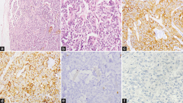
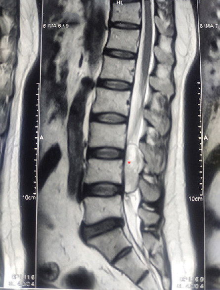
{"title":"Primary Carcinoid Tumor of the Central Nervous System: A Rare Case Report with a Diagnostic Challenge.","authors":"Anurag Singh, Alka Singh, Kamlesh Singh Bhaisora, Narendra Krishnani","doi":"10.4103/ijabmr.ijabmr_523_24","DOIUrl":null,"url":null,"abstract":"<p><p>Carcinoid tumors (CTs) are slow-growing neuroendocrine neoplasms that may arise in any part of the body. They usually affect the lungs or gut. Primary intradural extramedullary CTs of the central nervous system are rare; few cases have been reported. This case report describes a 36-year-old patient with lower back discomfort, weakness, and lower leg pain for 8 months. Contrast-enhanced magnetic resonance imaging of the lumbosacral spine, from the lower L3 vertebral level to the L4-5 disc level, revealed a 4.5 cm × 2.5 cm × 1.5 cm intradural mass lesion. The main differential diagnoses were on clinical and radiographic examination: nerve sheath tumors, meningiomas, and myxopapillary ependymomas. The tumor was excised en bloc after an L3-L5 lumbar laminectomy. Histomorphology and immunohistochemistry made a definitive diagnosis of CT of the lumbar spine. Two years after surgery, there was no clinical or radiological evidence of tumor recurrence or metastasis. The present case study is intended to effectively diagnose and treat spinal intradural extramedullary CTs.</p>","PeriodicalId":13727,"journal":{"name":"International Journal of Applied and Basic Medical Research","volume":"15 2","pages":"128-131"},"PeriodicalIF":0.8000,"publicationDate":"2025-04-01","publicationTypes":"Journal Article","fieldsOfStudy":null,"isOpenAccess":false,"openAccessPdf":"https://www.ncbi.nlm.nih.gov/pmc/articles/PMC12058044/pdf/","citationCount":"0","resultStr":null,"platform":"Semanticscholar","paperid":null,"PeriodicalName":"International Journal of Applied and Basic Medical Research","FirstCategoryId":"1085","ListUrlMain":"https://doi.org/10.4103/ijabmr.ijabmr_523_24","RegionNum":0,"RegionCategory":null,"ArticlePicture":[],"TitleCN":null,"AbstractTextCN":null,"PMCID":null,"EPubDate":"2025/4/7 0:00:00","PubModel":"Epub","JCR":"Q3","JCRName":"MEDICINE, GENERAL & INTERNAL","Score":null,"Total":0}
引用次数: 0
Abstract
Carcinoid tumors (CTs) are slow-growing neuroendocrine neoplasms that may arise in any part of the body. They usually affect the lungs or gut. Primary intradural extramedullary CTs of the central nervous system are rare; few cases have been reported. This case report describes a 36-year-old patient with lower back discomfort, weakness, and lower leg pain for 8 months. Contrast-enhanced magnetic resonance imaging of the lumbosacral spine, from the lower L3 vertebral level to the L4-5 disc level, revealed a 4.5 cm × 2.5 cm × 1.5 cm intradural mass lesion. The main differential diagnoses were on clinical and radiographic examination: nerve sheath tumors, meningiomas, and myxopapillary ependymomas. The tumor was excised en bloc after an L3-L5 lumbar laminectomy. Histomorphology and immunohistochemistry made a definitive diagnosis of CT of the lumbar spine. Two years after surgery, there was no clinical or radiological evidence of tumor recurrence or metastasis. The present case study is intended to effectively diagnose and treat spinal intradural extramedullary CTs.
类癌肿瘤(CTs)是生长缓慢的神经内分泌肿瘤,可出现在身体的任何部位。它们通常会影响肺部或肠道。原发性中枢神经系统硬膜内髓外ct是罕见的;报告的病例很少。本病例报告描述了一名36岁的患者,腰背部不适,无力和下肢疼痛8个月。腰骶棘磁共振增强成像,从下L3椎体水平到L4-5椎间盘水平,显示4.5 cm × 2.5 cm × 1.5 cm硬膜内肿块病变。主要鉴别诊断为临床及影像学检查:神经鞘瘤、脑膜瘤、黏液乳头状室管膜瘤。肿瘤在L3-L5腰椎椎板切除术后整体切除。组织形态学和免疫组织化学对腰椎的CT进行了明确的诊断。手术后两年,没有临床或放射学证据表明肿瘤复发或转移。本病例研究旨在有效诊断和治疗脊髓硬膜内髓外ct。

 求助内容:
求助内容: 应助结果提醒方式:


