Metformin induces mitochondria-mediated and endoplasmic reticulum stress-mediated apoptosis and inhibits angiogenesis-related gene expression in breast cancer cells via targeting VEGF-A/VEGFR2/NRP1.
Ares Alizade, Gulsah Evyapan, Ibrahim Seyfettin Celik, Berna Ozdem
{"title":"Metformin induces mitochondria-mediated and endoplasmic reticulum stress-mediated apoptosis and inhibits angiogenesis-related gene expression in breast cancer cells via targeting VEGF-A/VEGFR2/NRP1.","authors":"Ares Alizade, Gulsah Evyapan, Ibrahim Seyfettin Celik, Berna Ozdem","doi":"","DOIUrl":null,"url":null,"abstract":"<p><strong>Aim: </strong>To investigate the apoptotic and anti-angiogenic effects of metformin in human MCF7 breast cancer cells.</p><p><strong>Methods: </strong>The effect of metformin on cell viability was assessed by MTS and crystal violet assays, and its effect on cell migration was evaluated by the wound healing assay. The gene expression and protein levels of angiogenesis- and apoptosis-related genes were determined by real-time polymerase chain reaction, Western blot, and flow cytometry.</p><p><strong>Results: </strong>Metformin reduced the viability and migration of breast cancer cells compared with the control group. Furthermore, metformin (10 μM) increased the apoptosis-related gene and protein expression of caspase-3, Bax, AIF, CHOP and GRP78 48 hours after treatment compared with the control group. In contrast, it significantly decreased Bcl-2 and Wee1 gene and protein expression and suppressed angiogenesis-related genes VEGFA, VEGFR2, and NRP1.</p><p><strong>Conclusions: </strong>Our results suggest that metformin treatment activates apoptosis pathways and inactivates the angiogenesis pathway. Although this study was conducted in vitro and did not directly evaluate blood vessel formation, the observed downregulation of angiogenesis-related genes suggests potential anti-angiogenic activity of metformin at the gene expression level.</p>","PeriodicalId":10796,"journal":{"name":"Croatian Medical Journal","volume":"66 2","pages":"115-124"},"PeriodicalIF":2.3000,"publicationDate":"2025-05-07","publicationTypes":"Journal Article","fieldsOfStudy":null,"isOpenAccess":false,"openAccessPdf":"https://www.ncbi.nlm.nih.gov/pmc/articles/PMC12093123/pdf/","citationCount":"0","resultStr":null,"platform":"Semanticscholar","paperid":null,"PeriodicalName":"Croatian Medical Journal","FirstCategoryId":"3","ListUrlMain":"","RegionNum":4,"RegionCategory":"医学","ArticlePicture":[],"TitleCN":null,"AbstractTextCN":null,"PMCID":null,"EPubDate":"","PubModel":"","JCR":"Q2","JCRName":"MEDICINE, GENERAL & INTERNAL","Score":null,"Total":0}
引用次数: 0
Abstract
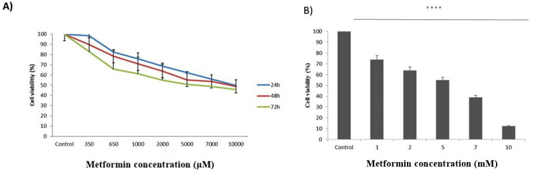
Aim: To investigate the apoptotic and anti-angiogenic effects of metformin in human MCF7 breast cancer cells.
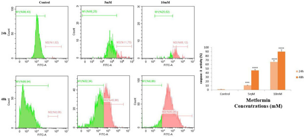
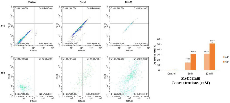
Methods: The effect of metformin on cell viability was assessed by MTS and crystal violet assays, and its effect on cell migration was evaluated by the wound healing assay. The gene expression and protein levels of angiogenesis- and apoptosis-related genes were determined by real-time polymerase chain reaction, Western blot, and flow cytometry.
Results: Metformin reduced the viability and migration of breast cancer cells compared with the control group. Furthermore, metformin (10 μM) increased the apoptosis-related gene and protein expression of caspase-3, Bax, AIF, CHOP and GRP78 48 hours after treatment compared with the control group. In contrast, it significantly decreased Bcl-2 and Wee1 gene and protein expression and suppressed angiogenesis-related genes VEGFA, VEGFR2, and NRP1.
Conclusions: Our results suggest that metformin treatment activates apoptosis pathways and inactivates the angiogenesis pathway. Although this study was conducted in vitro and did not directly evaluate blood vessel formation, the observed downregulation of angiogenesis-related genes suggests potential anti-angiogenic activity of metformin at the gene expression level.
期刊介绍:
Croatian Medical Journal (CMJ) is an international peer reviewed journal open to scientists from all fields of biomedicine and health related research.
Although CMJ welcomes all contributions that increase and expand on medical knowledge, the two areas are of the special interest: topics globally relevant for biomedicine and health and medicine in developing and emerging countries.

 求助内容:
求助内容: 应助结果提醒方式:


