Xuefeng Hou, Kun Chen, Huiwen Luo, Wengui Xu, Xiaofeng Li
{"title":"Identification of HER2-over-expression, HER2-low-expression, and HER2-zero-expression statuses in breast cancer based on <sup>18</sup>F-FDG PET/CT radiomics.","authors":"Xuefeng Hou, Kun Chen, Huiwen Luo, Wengui Xu, Xiaofeng Li","doi":"10.1186/s40644-025-00880-2","DOIUrl":null,"url":null,"abstract":"<p><strong>Purpose: </strong>According to the updated classification system, human epidermal growth factor receptor 2 (HER2) expression statuses are divided into the following three groups: HER2-over-expression, HER2-low-expression, and HER2-zero-expression. HER2-negative expression was reclassified into HER2-low-expression and HER2-zero-expression. This study aimed to identify three different HER2 expression statuses for breast cancer (BC) patients using PET/CT radiomics and clinicopathological characteristics.</p><p><strong>Methods and materials: </strong>A total of 315 BC patients who met the inclusion and exclusion criteria from two institutions were retrospectively included. The patients in institution 1 were divided into the training set and the independent validation set according to the ratio of 7:3, and institution 2 was used as the external validation set. According to the results of pathological examination, all BC patients were divided into HER2-over-expression, HER2-low-expression, and HER2-zero-expression. First, PET/CT radiomic features and clinicopathological features based on each patient were extracted and collected. Second, multiple methods were used to perform feature screening and feature selection. Then, four machine learning classifiers, including logistic regression (LR), k-nearest neighbor (KNN), support vector machine (SVM), and random forest (RF), were constructed to identify HER2-over-expression vs. others, HER2-low-expression vs. others, and HER2-zero-expression vs. others. The receiver operator characteristic (ROC) curve was plotted to measure the model's predictive power.</p><p><strong>Results: </strong>According to the feature screening process, 8, 10, and 2 radiomics features and 2 clinicopathological features were finally selected to construct three prediction models (HER2-over-expression vs. others, HER2-low-expression vs. others, and HER2-zero-expression vs. others). For HER2-over-expression vs. others, the RF model outperformed other models with an AUC value of 0.843 (95%CI: 0.774-0.897), 0.785 (95%CI: 0.665-0.877), and 0.788 (95%CI: 0.708-0.868) in the training set, independent validation set, and external validation set. Concerning HER2-low-expression vs. others, the outperformance of the LR model over other models was identified with an AUC value of 0.783 (95%CI: 0.708-0.846), 0.756 (95%CI: 0.634-0.854), and 0.779 (95%CI: 0.698-0.860) in the training set, independent validation set, and external validation set. Whereas, the KNN model was confirmed as the optimal model to distinguish HER2-zero-expression from others, with an AUC value of 0.929 (95%CI: 0.890-0.958), 0.847 (95%CI: 0.764-0.910), and 0.835 (95%CI: 0.762-0.908) in the training set, independent validation set, and external validation set.</p><p><strong>Conclusion: </strong>Combined PET/CT radiomic models integrating with clinicopathological characteristics are non-invasively predictive of different HER2 statuses of BC patients.</p>","PeriodicalId":9548,"journal":{"name":"Cancer Imaging","volume":"25 1","pages":"62"},"PeriodicalIF":3.5000,"publicationDate":"2025-05-12","publicationTypes":"Journal Article","fieldsOfStudy":null,"isOpenAccess":false,"openAccessPdf":"https://www.ncbi.nlm.nih.gov/pmc/articles/PMC12070556/pdf/","citationCount":"0","resultStr":null,"platform":"Semanticscholar","paperid":null,"PeriodicalName":"Cancer Imaging","FirstCategoryId":"3","ListUrlMain":"https://doi.org/10.1186/s40644-025-00880-2","RegionNum":2,"RegionCategory":"医学","ArticlePicture":[],"TitleCN":null,"AbstractTextCN":null,"PMCID":null,"EPubDate":"","PubModel":"","JCR":"Q2","JCRName":"ONCOLOGY","Score":null,"Total":0}
引用次数: 0
Abstract
Purpose: According to the updated classification system, human epidermal growth factor receptor 2 (HER2) expression statuses are divided into the following three groups: HER2-over-expression, HER2-low-expression, and HER2-zero-expression. HER2-negative expression was reclassified into HER2-low-expression and HER2-zero-expression. This study aimed to identify three different HER2 expression statuses for breast cancer (BC) patients using PET/CT radiomics and clinicopathological characteristics.
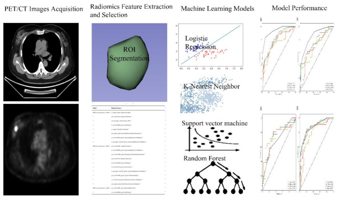
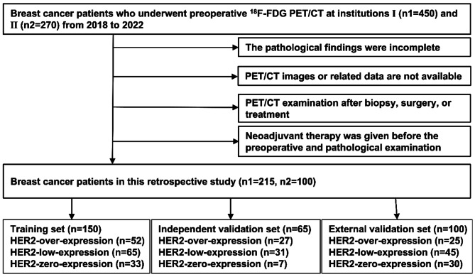
Methods and materials: A total of 315 BC patients who met the inclusion and exclusion criteria from two institutions were retrospectively included. The patients in institution 1 were divided into the training set and the independent validation set according to the ratio of 7:3, and institution 2 was used as the external validation set. According to the results of pathological examination, all BC patients were divided into HER2-over-expression, HER2-low-expression, and HER2-zero-expression. First, PET/CT radiomic features and clinicopathological features based on each patient were extracted and collected. Second, multiple methods were used to perform feature screening and feature selection. Then, four machine learning classifiers, including logistic regression (LR), k-nearest neighbor (KNN), support vector machine (SVM), and random forest (RF), were constructed to identify HER2-over-expression vs. others, HER2-low-expression vs. others, and HER2-zero-expression vs. others. The receiver operator characteristic (ROC) curve was plotted to measure the model's predictive power.
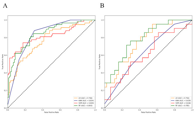
Results: According to the feature screening process, 8, 10, and 2 radiomics features and 2 clinicopathological features were finally selected to construct three prediction models (HER2-over-expression vs. others, HER2-low-expression vs. others, and HER2-zero-expression vs. others). For HER2-over-expression vs. others, the RF model outperformed other models with an AUC value of 0.843 (95%CI: 0.774-0.897), 0.785 (95%CI: 0.665-0.877), and 0.788 (95%CI: 0.708-0.868) in the training set, independent validation set, and external validation set. Concerning HER2-low-expression vs. others, the outperformance of the LR model over other models was identified with an AUC value of 0.783 (95%CI: 0.708-0.846), 0.756 (95%CI: 0.634-0.854), and 0.779 (95%CI: 0.698-0.860) in the training set, independent validation set, and external validation set. Whereas, the KNN model was confirmed as the optimal model to distinguish HER2-zero-expression from others, with an AUC value of 0.929 (95%CI: 0.890-0.958), 0.847 (95%CI: 0.764-0.910), and 0.835 (95%CI: 0.762-0.908) in the training set, independent validation set, and external validation set.
Conclusion: Combined PET/CT radiomic models integrating with clinicopathological characteristics are non-invasively predictive of different HER2 statuses of BC patients.
Cancer ImagingONCOLOGY-RADIOLOGY, NUCLEAR MEDICINE & MEDICAL IMAGING
CiteScore
7.00
自引率
0.00%
发文量
66
审稿时长
>12 weeks
期刊介绍:
Cancer Imaging is an open access, peer-reviewed journal publishing original articles, reviews and editorials written by expert international radiologists working in oncology.
The journal encompasses CT, MR, PET, ultrasound, radionuclide and multimodal imaging in all kinds of malignant tumours, plus new developments, techniques and innovations. Topics of interest include:
Breast Imaging
Chest
Complications of treatment
Ear, Nose & Throat
Gastrointestinal
Hepatobiliary & Pancreatic
Imaging biomarkers
Interventional
Lymphoma
Measurement of tumour response
Molecular functional imaging
Musculoskeletal
Neuro oncology
Nuclear Medicine
Paediatric.

 求助内容:
求助内容: 应助结果提醒方式:


