Christina L Greene, Geoffrey Traeger, Akshay Venkatesh, David Han, Mark W Majesky
{"title":"Origins of Aortic Coarctation: A Vascular Smooth Muscle Compartment Boundary Model.","authors":"Christina L Greene, Geoffrey Traeger, Akshay Venkatesh, David Han, Mark W Majesky","doi":"10.3390/jdb13020013","DOIUrl":null,"url":null,"abstract":"<p><p>Compartment boundaries divide the embryo into segments with distinct fates and functions. In the vascular system, compartment boundaries organize endothelial cells into arteries, capillaries, and veins that are the fundamental units of a circulatory network. For vascular smooth muscle cells (SMCs), such boundaries produce mosaic patterns of investment based on embryonic origins with important implications for the non-uniform distribution of vascular disease later in life. The morphogenesis of blood vessels requires vascular cell movements within compartments as highly-sensitive responses to changes in fluid flow shear stress and wall strain. These movements underline the remodeling of primitive plexuses, expansion of lumen diameters, regression of unused vessels, and building of multilayered artery walls. Although the loss of endothelial compartment boundaries can produce arterial-venous malformations, little is known about the consequences of mislocalization or the failure to form SMC-origin-specific boundaries during vascular development. We propose that the failure to establish a normal compartment boundary between cardiac neural-crest-derived SMCs of the 6th pharyngeal arch artery (future ductus arteriosus) and paraxial-mesoderm-derived SMCs of the dorsal aorta in mid-gestation embryos leads to aortic coarctation observed at birth. This model raises new questions about the effects of fluid flow dynamics on SMC investment and the formation of SMC compartment borders during pharyngeal arch artery remodeling and vascular development.</p>","PeriodicalId":15563,"journal":{"name":"Journal of Developmental Biology","volume":"13 2","pages":""},"PeriodicalIF":2.5000,"publicationDate":"2025-04-18","publicationTypes":"Journal Article","fieldsOfStudy":null,"isOpenAccess":false,"openAccessPdf":"https://www.ncbi.nlm.nih.gov/pmc/articles/PMC12015864/pdf/","citationCount":"0","resultStr":null,"platform":"Semanticscholar","paperid":null,"PeriodicalName":"Journal of Developmental Biology","FirstCategoryId":"1085","ListUrlMain":"https://doi.org/10.3390/jdb13020013","RegionNum":0,"RegionCategory":null,"ArticlePicture":[],"TitleCN":null,"AbstractTextCN":null,"PMCID":null,"EPubDate":"","PubModel":"","JCR":"Q3","JCRName":"DEVELOPMENTAL BIOLOGY","Score":null,"Total":0}
引用次数: 0
Abstract
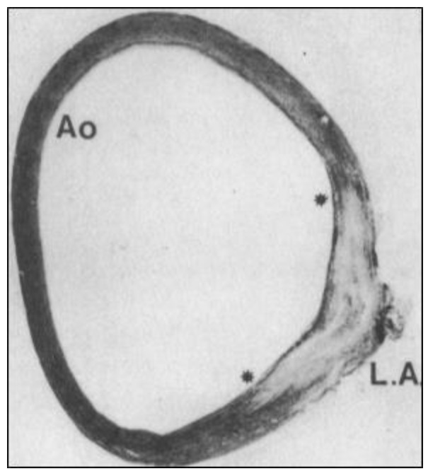
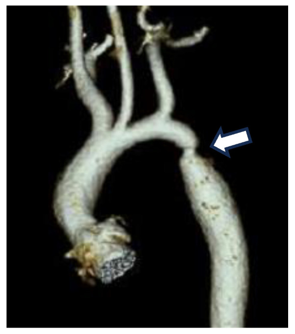
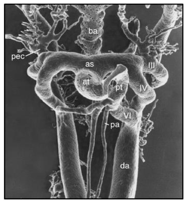
Compartment boundaries divide the embryo into segments with distinct fates and functions. In the vascular system, compartment boundaries organize endothelial cells into arteries, capillaries, and veins that are the fundamental units of a circulatory network. For vascular smooth muscle cells (SMCs), such boundaries produce mosaic patterns of investment based on embryonic origins with important implications for the non-uniform distribution of vascular disease later in life. The morphogenesis of blood vessels requires vascular cell movements within compartments as highly-sensitive responses to changes in fluid flow shear stress and wall strain. These movements underline the remodeling of primitive plexuses, expansion of lumen diameters, regression of unused vessels, and building of multilayered artery walls. Although the loss of endothelial compartment boundaries can produce arterial-venous malformations, little is known about the consequences of mislocalization or the failure to form SMC-origin-specific boundaries during vascular development. We propose that the failure to establish a normal compartment boundary between cardiac neural-crest-derived SMCs of the 6th pharyngeal arch artery (future ductus arteriosus) and paraxial-mesoderm-derived SMCs of the dorsal aorta in mid-gestation embryos leads to aortic coarctation observed at birth. This model raises new questions about the effects of fluid flow dynamics on SMC investment and the formation of SMC compartment borders during pharyngeal arch artery remodeling and vascular development.
期刊介绍:
The Journal of Developmental Biology (ISSN 2221-3759) is an international, peer-reviewed, quick-refereeing, open access journal, which publishes reviews, research papers and communications on the development of multicellular organisms at the molecule, cell, tissue, organ and whole organism levels. Our aim is to encourage researchers to effortlessly publish their new findings or concepts rapidly in an open access medium, overseen by their peers. There is no restriction on the length of the papers; the full experimental details must be provided so that the results can be reproduced. Electronic files regarding the full details of the experimental procedure, if unable to be published in a normal way, can be deposited as supplementary material. Journal of Developmental Biology focuses on: -Development mechanisms and genetics -Cell differentiation -Embryonal development -Tissue/organism growth -Metamorphosis and regeneration of the organisms. It involves many biological fields, such as Molecular biology, Genetics, Physiology, Cell biology, Anatomy, Embryology, Cancer research, Neurobiology, Immunology, Ecology, Evolutionary biology.

 求助内容:
求助内容: 应助结果提醒方式:


