Rawan Hosny, Jylan Gouda, Tamer A Macky, Ayman Khattab, Hany Mekkawy, Abdussalam M Abdullatif
{"title":"OCT macular changes in type 1 ROP following Ranibizumab injections.","authors":"Rawan Hosny, Jylan Gouda, Tamer A Macky, Ayman Khattab, Hany Mekkawy, Abdussalam M Abdullatif","doi":"10.1186/s40942-025-00664-7","DOIUrl":null,"url":null,"abstract":"<p><strong>Aim: </strong>To investigate the OCT macular changes in type 1 ROP one month following Ranibizumab injections.</p><p><strong>Methods: </strong>Preterm infants with type 1 ROP indicated for Ranibizumab injections were included in this study. Handheld OCT imaging was performed at baseline, 1 week, and 1 month post injection. Central full thickness (CFT), inner retinal layer (IRL), and outer retinal layer (ORL) thickness measurements were taken from foveal center and parafoveal region.</p><p><strong>Results: </strong>24 eyes of 12 infants were included in this study. There were no significant changes in the mean CFT and IRL thickness at 1 month (p = 0.5 and 0.1 respectively). However, there was significant increase in the mean ORL thickness at 1 month (69.9 ± 16, 96.1 ± 25 at baseline and one month respectively, p < 0.001), with differentiation (appearance of IS/OS junction ± ELM) in 55.6% of eyes. Macular edema (ME) was observed in 12 eyes (50%) and was associated with smaller birth weight (p = 0.0290). There was no significant decrease in mean CFT in eyes with ME at 1 month (p = 0.13), with complete resolution in only 6 eyes (50%) during the study period. Regression of plus was associated with lower CFT (1 week and 1 month; p = 0.02 and 0.03, respectively).</p><p><strong>Conclusion: </strong>Ranibizumab treated eyes in type 1 show ORL thickening and differentiation but with inadequate resolution of ME.</p>","PeriodicalId":14289,"journal":{"name":"International Journal of Retina and Vitreous","volume":"11 1","pages":"47"},"PeriodicalIF":2.4000,"publicationDate":"2025-04-17","publicationTypes":"Journal Article","fieldsOfStudy":null,"isOpenAccess":false,"openAccessPdf":"https://www.ncbi.nlm.nih.gov/pmc/articles/PMC12004622/pdf/","citationCount":"0","resultStr":null,"platform":"Semanticscholar","paperid":null,"PeriodicalName":"International Journal of Retina and Vitreous","FirstCategoryId":"1085","ListUrlMain":"https://doi.org/10.1186/s40942-025-00664-7","RegionNum":0,"RegionCategory":null,"ArticlePicture":[],"TitleCN":null,"AbstractTextCN":null,"PMCID":null,"EPubDate":"","PubModel":"","JCR":"Q2","JCRName":"OPHTHALMOLOGY","Score":null,"Total":0}
引用次数: 0
Abstract
Aim: To investigate the OCT macular changes in type 1 ROP one month following Ranibizumab injections.
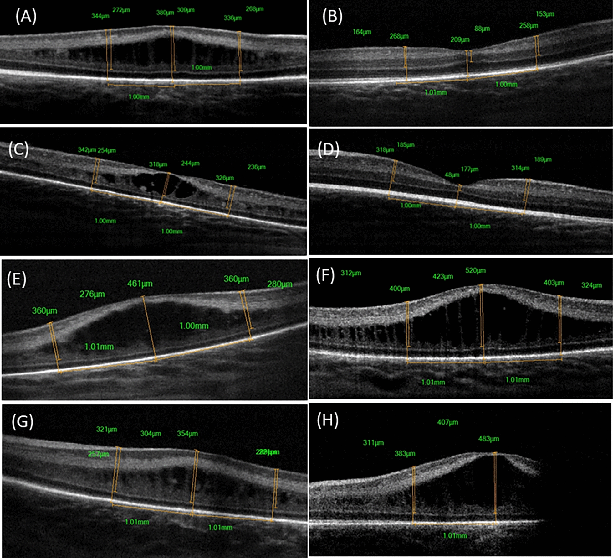
Methods: Preterm infants with type 1 ROP indicated for Ranibizumab injections were included in this study. Handheld OCT imaging was performed at baseline, 1 week, and 1 month post injection. Central full thickness (CFT), inner retinal layer (IRL), and outer retinal layer (ORL) thickness measurements were taken from foveal center and parafoveal region.
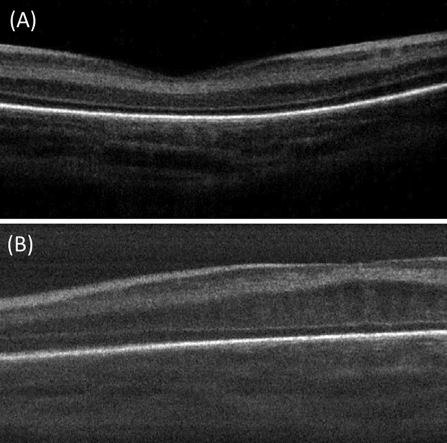
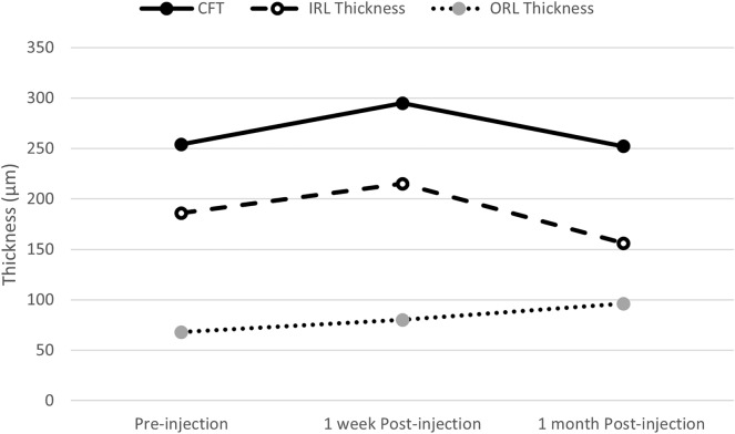
Results: 24 eyes of 12 infants were included in this study. There were no significant changes in the mean CFT and IRL thickness at 1 month (p = 0.5 and 0.1 respectively). However, there was significant increase in the mean ORL thickness at 1 month (69.9 ± 16, 96.1 ± 25 at baseline and one month respectively, p < 0.001), with differentiation (appearance of IS/OS junction ± ELM) in 55.6% of eyes. Macular edema (ME) was observed in 12 eyes (50%) and was associated with smaller birth weight (p = 0.0290). There was no significant decrease in mean CFT in eyes with ME at 1 month (p = 0.13), with complete resolution in only 6 eyes (50%) during the study period. Regression of plus was associated with lower CFT (1 week and 1 month; p = 0.02 and 0.03, respectively).
Conclusion: Ranibizumab treated eyes in type 1 show ORL thickening and differentiation but with inadequate resolution of ME.
期刊介绍:
International Journal of Retina and Vitreous focuses on the ophthalmic subspecialty of vitreoretinal disorders. The journal presents original articles on new approaches to diagnosis, outcomes of clinical trials, innovations in pharmacological therapy and surgical techniques, as well as basic science advances that impact clinical practice. Topical areas include, but are not limited to: -Imaging of the retina, choroid and vitreous -Innovations in optical coherence tomography (OCT) -Small-gauge vitrectomy, retinal detachment, chromovitrectomy -Electroretinography (ERG), microperimetry, other functional tests -Intraocular tumors -Retinal pharmacotherapy & drug delivery -Diabetic retinopathy & other vascular diseases -Age-related macular degeneration (AMD) & other macular entities

 求助内容:
求助内容: 应助结果提醒方式:


