Andreas Heinrich, Michael Hubig, Gita Mall, Ulf Teichgräber
{"title":"Computer vision-based personal identification using 2D maximum intensity projection CT images.","authors":"Andreas Heinrich, Michael Hubig, Gita Mall, Ulf Teichgräber","doi":"10.1007/s00330-025-11630-0","DOIUrl":null,"url":null,"abstract":"<p><strong>Objectives: </strong>Computer vision (CV) mimics human vision, enabling the automatic comparison of radiological images from recent examinations with a vast image database for unique identification. This method offers significant potential in emergencies involving unknown individuals. This study assesses whether maximum intensity projection (MIP) images from thoracic computed tomography (CT) examinations are suitable for automated CV-based personal identification.</p><p><strong>Methods: </strong>The study analyzed 12,465 native CT examinations of the thorax from 8177 individuals, focusing on MIP images to assess their potential for CV-based personal identification in 300 cases. CV automatically identifies and describes features in images, which are then matched to reference images. The number of matching points was used as an indicator of identification accuracy.</p><p><strong>Results: </strong>The identification rate was 98.67% (296/300) at rank 1 and 99.67% (299/300) at rank 10, among over 8177 potential identities. Matching points were higher for images of the same individual (7.43 ± 5.83%) compared to different individuals (0.16 ± 0.14%), with 100% representing the maximum possible matching points. Reliable matching points were mainly found in the thoracic skeleton, sternum, and spine. Challenges arose when the patient was curved on the table or when medical equipment was present in the image.</p><p><strong>Conclusion: </strong>Unambiguous identification based on MIP images from thoracic CT examinations is highly reliable, even for large CV databases. This method is applicable to various 2D reconstructions, provided anatomical structures are comparably represented. Radiology offers extensive reference images for CV databases, enhancing automated personal identification in emergencies.</p><p><strong>Key points: </strong>Question Computer vision-based personal identification holds great potential, but it remains unclear whether maximum intensity projection images from thoracic-CT scans are suitable for this purpose. Findings Maximum intensity projection images of the thorax are highly individual, with computer vision-based identification achieving nearly 100% rank-1 accuracy across a potential 8177 identities. Clinical relevance Radiology holds a vast collection of reference images for a computer vision database, enabling automated personal identification in emergency examinations. This improves patient care and communication with relatives by providing access to medical history.</p>","PeriodicalId":12076,"journal":{"name":"European Radiology","volume":" ","pages":"5900-5910"},"PeriodicalIF":4.7000,"publicationDate":"2025-10-01","publicationTypes":"Journal Article","fieldsOfStudy":null,"isOpenAccess":false,"openAccessPdf":"https://www.ncbi.nlm.nih.gov/pmc/articles/PMC12417270/pdf/","citationCount":"0","resultStr":null,"platform":"Semanticscholar","paperid":null,"PeriodicalName":"European Radiology","FirstCategoryId":"3","ListUrlMain":"https://doi.org/10.1007/s00330-025-11630-0","RegionNum":2,"RegionCategory":"医学","ArticlePicture":[],"TitleCN":null,"AbstractTextCN":null,"PMCID":null,"EPubDate":"2025/4/27 0:00:00","PubModel":"Epub","JCR":"Q1","JCRName":"RADIOLOGY, NUCLEAR MEDICINE & MEDICAL IMAGING","Score":null,"Total":0}
引用次数: 0
Abstract
Objectives: Computer vision (CV) mimics human vision, enabling the automatic comparison of radiological images from recent examinations with a vast image database for unique identification. This method offers significant potential in emergencies involving unknown individuals. This study assesses whether maximum intensity projection (MIP) images from thoracic computed tomography (CT) examinations are suitable for automated CV-based personal identification.
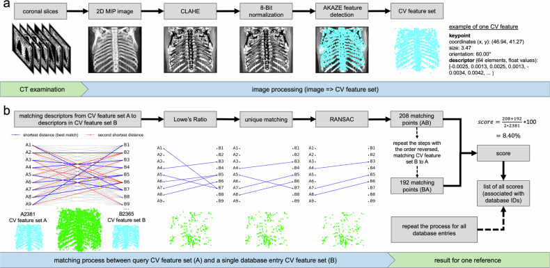
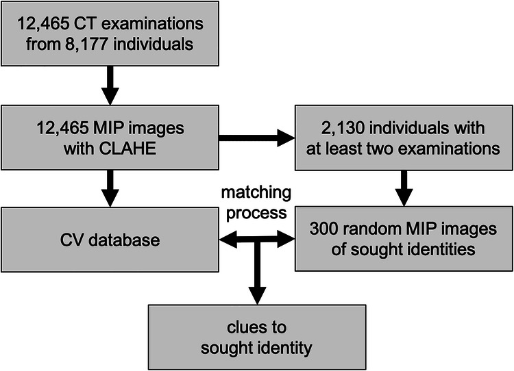
Methods: The study analyzed 12,465 native CT examinations of the thorax from 8177 individuals, focusing on MIP images to assess their potential for CV-based personal identification in 300 cases. CV automatically identifies and describes features in images, which are then matched to reference images. The number of matching points was used as an indicator of identification accuracy.
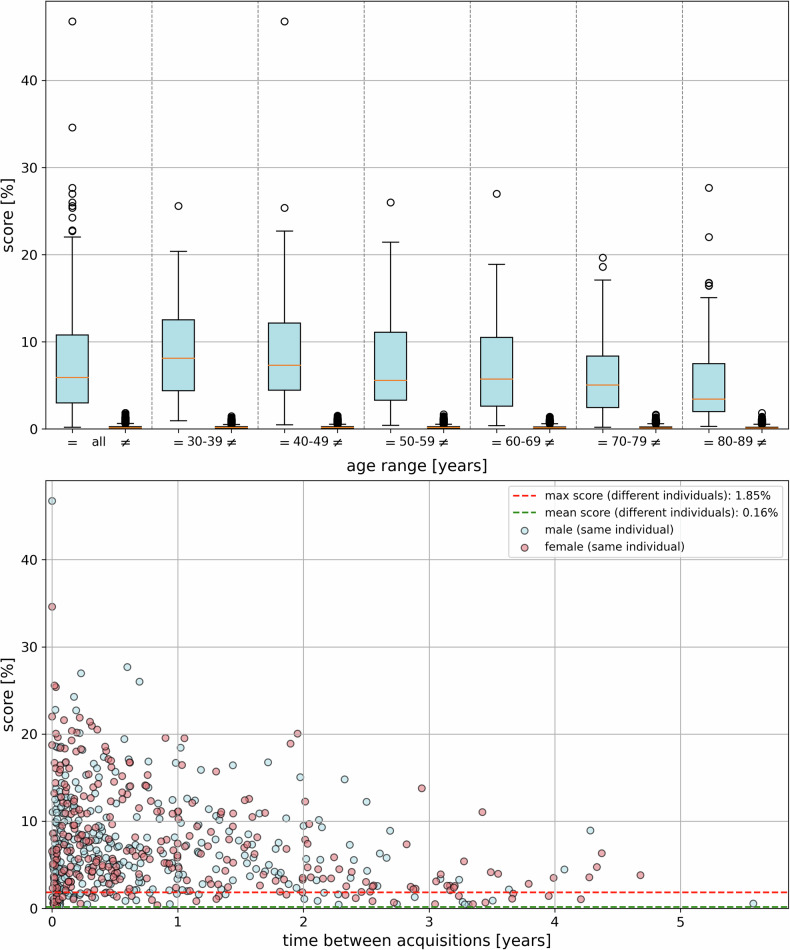
Results: The identification rate was 98.67% (296/300) at rank 1 and 99.67% (299/300) at rank 10, among over 8177 potential identities. Matching points were higher for images of the same individual (7.43 ± 5.83%) compared to different individuals (0.16 ± 0.14%), with 100% representing the maximum possible matching points. Reliable matching points were mainly found in the thoracic skeleton, sternum, and spine. Challenges arose when the patient was curved on the table or when medical equipment was present in the image.
Conclusion: Unambiguous identification based on MIP images from thoracic CT examinations is highly reliable, even for large CV databases. This method is applicable to various 2D reconstructions, provided anatomical structures are comparably represented. Radiology offers extensive reference images for CV databases, enhancing automated personal identification in emergencies.
Key points: Question Computer vision-based personal identification holds great potential, but it remains unclear whether maximum intensity projection images from thoracic-CT scans are suitable for this purpose. Findings Maximum intensity projection images of the thorax are highly individual, with computer vision-based identification achieving nearly 100% rank-1 accuracy across a potential 8177 identities. Clinical relevance Radiology holds a vast collection of reference images for a computer vision database, enabling automated personal identification in emergency examinations. This improves patient care and communication with relatives by providing access to medical history.
期刊介绍:
European Radiology (ER) continuously updates scientific knowledge in radiology by publication of strong original articles and state-of-the-art reviews written by leading radiologists. A well balanced combination of review articles, original papers, short communications from European radiological congresses and information on society matters makes ER an indispensable source for current information in this field.
This is the Journal of the European Society of Radiology, and the official journal of a number of societies.
From 2004-2008 supplements to European Radiology were published under its companion, European Radiology Supplements, ISSN 1613-3749.

 求助内容:
求助内容: 应助结果提醒方式:


