{"title":"The impact of a simple positioning aid device on the diagnostic performance of thyroid cancer in CT scans: a randomized controlled trial.","authors":"Wei-Hua Lin, Hui-Juan Huang, Wen-Cong Yang, Qing-Wen Huang, Rui-Gang Huang, Fu-Rong Luo, Dong-Yi Chen, Zheng-Han Yang, Hai-Tao Li, Hui-Huang Zeng, Hui-Jun Xiao","doi":"10.1186/s40644-025-00878-w","DOIUrl":null,"url":null,"abstract":"<p><strong>Objective: </strong>To evaluate the effectiveness of a simple positioning aid device in neck CT scans for the diagnosis of thyroid cancer, with a focus on its influence on image quality and diagnostic accuracy.</p><p><strong>Methods: </strong>A randomized controlled trial was conducted involving 180 patients with suspected thyroid cancer. Participants were randomly assigned to two groups: the device-assisted positioning group (Group A) and the traditional positioning group (Group B). A total of 147 patients who underwent enhanced neck CT scans and subsequent surgical pathological biopsies were included in the final analysis. Image quality and thyroid disease diagnoses were independently assessed by two experienced radiologists, with a unified consensus for the final conclusions. Objective imaging parameters and subjective ratings were used to evaluate image quality. Pathological findings served as the gold standard to compare the diagnostic accuracy of the two groups for thyroid malignancy, capsular invasion, and lymph node metastasis. Additionally, radiation doses in both groups were compared.</p><p><strong>Results: </strong>A total of 147 patients were included in the analysis, with 72 patients in Group A and 75 in Group B. The baseline characteristics of the two groups were similar (P > 0.05). Group A demonstrated significantly superior image quality compared to Group B, with shorter length of artifacts (LA), lower proportion of affected thyroid (PA), and lower artifact index (AI). Subjective assessments also favored Group A, showing better ratings for regional artifacts and overall image quality. In terms of diagnostic accuracy, Group A outperformed Group B in detecting thyroid cancer (AUC: 0.852 vs. 0.676, P = 0.021). For the right thyroid lobe, Group A had significantly better diagnostic performance (AUC: 0.897 vs. 0.746, P = 0.016). Group A also showed superior performance in diagnosing capsular invasion (AUC: 0.861 vs. 0.721, P = 0.037), with similar results observed for both the left and right thyroid lobes. There was no significant difference between the groups in diagnosing lymph node metastasis. Furthermore, thyroid region radiation doses (CTDIvol and SSDE) were significantly lower in Group A compared to Group B.</p><p><strong>Conclusion: </strong>The use of a positioning aid device significantly improves CT image quality, enhancing diagnostic accuracy for malignant thyroid lesions and capsular invasion, while also reducing radiation exposure.</p>","PeriodicalId":9548,"journal":{"name":"Cancer Imaging","volume":"25 1","pages":"60"},"PeriodicalIF":3.5000,"publicationDate":"2025-05-08","publicationTypes":"Journal Article","fieldsOfStudy":null,"isOpenAccess":false,"openAccessPdf":"https://www.ncbi.nlm.nih.gov/pmc/articles/PMC12063306/pdf/","citationCount":"0","resultStr":null,"platform":"Semanticscholar","paperid":null,"PeriodicalName":"Cancer Imaging","FirstCategoryId":"3","ListUrlMain":"https://doi.org/10.1186/s40644-025-00878-w","RegionNum":2,"RegionCategory":"医学","ArticlePicture":[],"TitleCN":null,"AbstractTextCN":null,"PMCID":null,"EPubDate":"","PubModel":"","JCR":"Q2","JCRName":"ONCOLOGY","Score":null,"Total":0}
引用次数: 0
Abstract
Objective: To evaluate the effectiveness of a simple positioning aid device in neck CT scans for the diagnosis of thyroid cancer, with a focus on its influence on image quality and diagnostic accuracy.
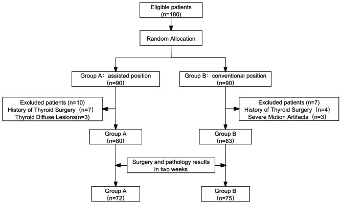
Methods: A randomized controlled trial was conducted involving 180 patients with suspected thyroid cancer. Participants were randomly assigned to two groups: the device-assisted positioning group (Group A) and the traditional positioning group (Group B). A total of 147 patients who underwent enhanced neck CT scans and subsequent surgical pathological biopsies were included in the final analysis. Image quality and thyroid disease diagnoses were independently assessed by two experienced radiologists, with a unified consensus for the final conclusions. Objective imaging parameters and subjective ratings were used to evaluate image quality. Pathological findings served as the gold standard to compare the diagnostic accuracy of the two groups for thyroid malignancy, capsular invasion, and lymph node metastasis. Additionally, radiation doses in both groups were compared.
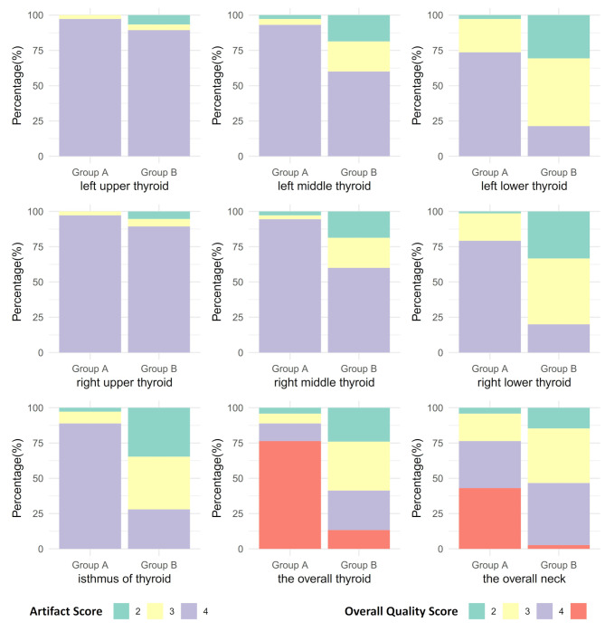
Results: A total of 147 patients were included in the analysis, with 72 patients in Group A and 75 in Group B. The baseline characteristics of the two groups were similar (P > 0.05). Group A demonstrated significantly superior image quality compared to Group B, with shorter length of artifacts (LA), lower proportion of affected thyroid (PA), and lower artifact index (AI). Subjective assessments also favored Group A, showing better ratings for regional artifacts and overall image quality. In terms of diagnostic accuracy, Group A outperformed Group B in detecting thyroid cancer (AUC: 0.852 vs. 0.676, P = 0.021). For the right thyroid lobe, Group A had significantly better diagnostic performance (AUC: 0.897 vs. 0.746, P = 0.016). Group A also showed superior performance in diagnosing capsular invasion (AUC: 0.861 vs. 0.721, P = 0.037), with similar results observed for both the left and right thyroid lobes. There was no significant difference between the groups in diagnosing lymph node metastasis. Furthermore, thyroid region radiation doses (CTDIvol and SSDE) were significantly lower in Group A compared to Group B.
Conclusion: The use of a positioning aid device significantly improves CT image quality, enhancing diagnostic accuracy for malignant thyroid lesions and capsular invasion, while also reducing radiation exposure.
Cancer ImagingONCOLOGY-RADIOLOGY, NUCLEAR MEDICINE & MEDICAL IMAGING
CiteScore
7.00
自引率
0.00%
发文量
66
审稿时长
>12 weeks
期刊介绍:
Cancer Imaging is an open access, peer-reviewed journal publishing original articles, reviews and editorials written by expert international radiologists working in oncology.
The journal encompasses CT, MR, PET, ultrasound, radionuclide and multimodal imaging in all kinds of malignant tumours, plus new developments, techniques and innovations. Topics of interest include:
Breast Imaging
Chest
Complications of treatment
Ear, Nose & Throat
Gastrointestinal
Hepatobiliary & Pancreatic
Imaging biomarkers
Interventional
Lymphoma
Measurement of tumour response
Molecular functional imaging
Musculoskeletal
Neuro oncology
Nuclear Medicine
Paediatric.

 求助内容:
求助内容: 应助结果提醒方式:


