Martine Remy-Jardin, Alain Duhamel, Marie Delobelle, Jean-François Bervar, Thomas Flohr, Jacques Remy
{"title":"Lung microvasculopathy in chronic thromboembolic pulmonary hypertension: high-resolution findings with photon-counting detector CT in 29 patients.","authors":"Martine Remy-Jardin, Alain Duhamel, Marie Delobelle, Jean-François Bervar, Thomas Flohr, Jacques Remy","doi":"10.1007/s00330-025-11561-w","DOIUrl":null,"url":null,"abstract":"<p><strong>Purpose: </strong>To evaluate CT findings suggestive of lung microvasculopathy in patients with chronic thromboembolic pulmonary hypertension (CTEPH).</p><p><strong>Materials and methods: </strong>Twenty-nine patients were scanned with high-spatial resolution on a photon-counting detector (PCD)-CT unit. A maximum of three pairs per patient, each composed of hyper- and hypo-attenuating areas of mosaic perfusion, were selected.</p><p><strong>Results: </strong>Comparative analysis of the 86 selected pairs showed: (a) a higher frequency of ill-defined micronodules (p = 0.008), lobular ground-glass opacities (p = 0.01) and haziness (p = 0.003) in hypoattenuated areas; (b) there was no significant difference in the frequency of neovascularity (p = 0.43). Similar trends were observed in hypoattenuating areas of the 66 pairs studied in the 22 patients with central and peripheral CTEPH; an absence of ill-defined micronodules, lobular ground-glass opacities, and haziness in hyperattenuating areas was noticed in the 20 pairs studied in the 7 patients with peripheral CTEPH. Patients with a mean pulmonary artery pressure ≤ 42 mmHg (i.e., the median value of mean pulmonary artery pressure) had 45 pairs compared, showing a higher frequency of ill-defined micronodules (p = 0.003) and haziness (p < 0.001) in hypoattenuated areas, together with a higher frequency of subpleural systemic-to-pulmonary anastomoses (p = 0.02). There were no statistical differences in the frequency of CT findings between hypo- and hyper-attenuating areas in the 41 pairs of patients with a mean pulmonary artery pressure > 42 mm Hg.</p><p><strong>Conclusion: </strong>CT features suggestive of microvasculopathy were more frequent in areas of hypoperfusion, with a trend toward homogenization of CT findings in patients with severe PH.</p><p><strong>Key points: </strong>Question Lung microvascular lesions play a crucial role in the origin of residual pulmonary hypertension after successful thromboendarterectomy, currently beyond the scope of imaging. Findings The expected morphological abnormalities at the level of distal pulmonary circulation in CTEPH were found to be depictable in each zone of mosaic perfusion. Clinical relevance This study suggests that the high-spatial resolution of PCD-CT has the capability of approaching the complex pathophysiology of small-vessel disease in CTEPH, providing important information prior to therapeutic decisions.</p>","PeriodicalId":12076,"journal":{"name":"European Radiology","volume":" ","pages":"6369-6381"},"PeriodicalIF":4.7000,"publicationDate":"2025-10-01","publicationTypes":"Journal Article","fieldsOfStudy":null,"isOpenAccess":false,"openAccessPdf":"https://www.ncbi.nlm.nih.gov/pmc/articles/PMC12417283/pdf/","citationCount":"0","resultStr":null,"platform":"Semanticscholar","paperid":null,"PeriodicalName":"European Radiology","FirstCategoryId":"3","ListUrlMain":"https://doi.org/10.1007/s00330-025-11561-w","RegionNum":2,"RegionCategory":"医学","ArticlePicture":[],"TitleCN":null,"AbstractTextCN":null,"PMCID":null,"EPubDate":"2025/4/18 0:00:00","PubModel":"Epub","JCR":"Q1","JCRName":"RADIOLOGY, NUCLEAR MEDICINE & MEDICAL IMAGING","Score":null,"Total":0}
引用次数: 0
Abstract
Purpose: To evaluate CT findings suggestive of lung microvasculopathy in patients with chronic thromboembolic pulmonary hypertension (CTEPH).
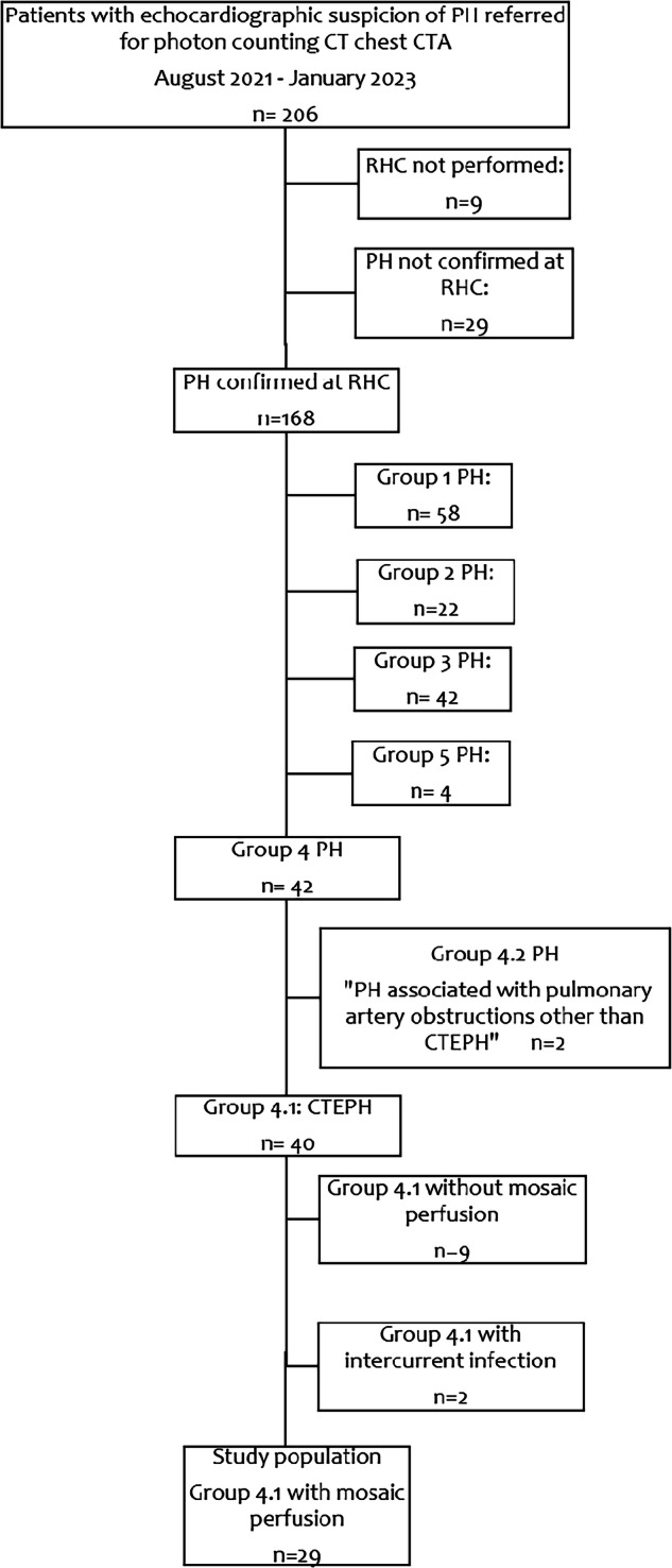
Materials and methods: Twenty-nine patients were scanned with high-spatial resolution on a photon-counting detector (PCD)-CT unit. A maximum of three pairs per patient, each composed of hyper- and hypo-attenuating areas of mosaic perfusion, were selected.
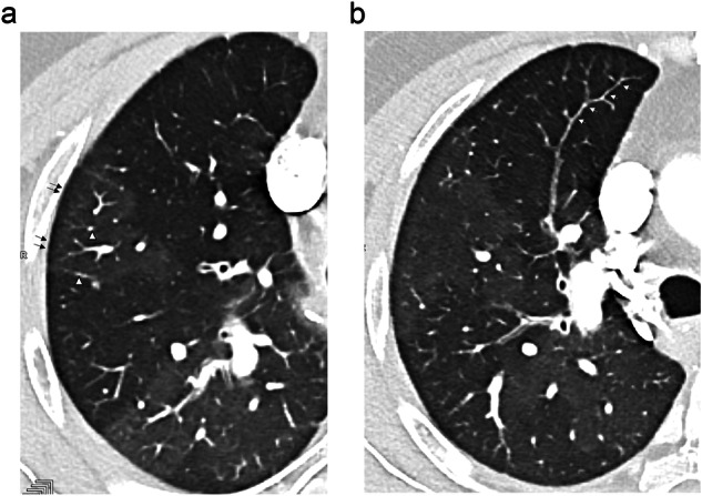
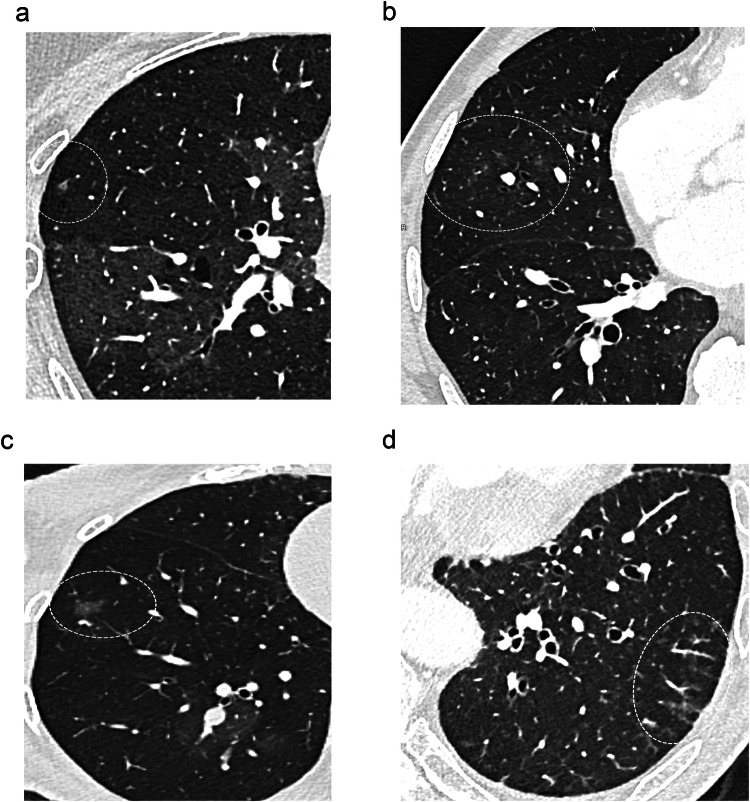
Results: Comparative analysis of the 86 selected pairs showed: (a) a higher frequency of ill-defined micronodules (p = 0.008), lobular ground-glass opacities (p = 0.01) and haziness (p = 0.003) in hypoattenuated areas; (b) there was no significant difference in the frequency of neovascularity (p = 0.43). Similar trends were observed in hypoattenuating areas of the 66 pairs studied in the 22 patients with central and peripheral CTEPH; an absence of ill-defined micronodules, lobular ground-glass opacities, and haziness in hyperattenuating areas was noticed in the 20 pairs studied in the 7 patients with peripheral CTEPH. Patients with a mean pulmonary artery pressure ≤ 42 mmHg (i.e., the median value of mean pulmonary artery pressure) had 45 pairs compared, showing a higher frequency of ill-defined micronodules (p = 0.003) and haziness (p < 0.001) in hypoattenuated areas, together with a higher frequency of subpleural systemic-to-pulmonary anastomoses (p = 0.02). There were no statistical differences in the frequency of CT findings between hypo- and hyper-attenuating areas in the 41 pairs of patients with a mean pulmonary artery pressure > 42 mm Hg.
Conclusion: CT features suggestive of microvasculopathy were more frequent in areas of hypoperfusion, with a trend toward homogenization of CT findings in patients with severe PH.
Key points: Question Lung microvascular lesions play a crucial role in the origin of residual pulmonary hypertension after successful thromboendarterectomy, currently beyond the scope of imaging. Findings The expected morphological abnormalities at the level of distal pulmonary circulation in CTEPH were found to be depictable in each zone of mosaic perfusion. Clinical relevance This study suggests that the high-spatial resolution of PCD-CT has the capability of approaching the complex pathophysiology of small-vessel disease in CTEPH, providing important information prior to therapeutic decisions.
期刊介绍:
European Radiology (ER) continuously updates scientific knowledge in radiology by publication of strong original articles and state-of-the-art reviews written by leading radiologists. A well balanced combination of review articles, original papers, short communications from European radiological congresses and information on society matters makes ER an indispensable source for current information in this field.
This is the Journal of the European Society of Radiology, and the official journal of a number of societies.
From 2004-2008 supplements to European Radiology were published under its companion, European Radiology Supplements, ISSN 1613-3749.

 求助内容:
求助内容: 应助结果提醒方式:


