Measurement of Twitch Dynamics in Response to Exercise Induced Changes in Mitochondrial Disease Using Motor Unit Magnetic Resonance Imaging (MUMRI): A Proof-of-Concept Study.
Matthew G Birkbeck, Mathew Elameer, Linda Heskamp, Jane Newman, Renae J Stefanetti, Isabel Barrow, Oksana Pogoryelova, Gráinne S Gorman, Julie Hall, Ian S Schofield, Andrew M Blamire, Roger G Whittaker
{"title":"Measurement of Twitch Dynamics in Response to Exercise Induced Changes in Mitochondrial Disease Using Motor Unit Magnetic Resonance Imaging (MUMRI): A Proof-of-Concept Study.","authors":"Matthew G Birkbeck, Mathew Elameer, Linda Heskamp, Jane Newman, Renae J Stefanetti, Isabel Barrow, Oksana Pogoryelova, Gráinne S Gorman, Julie Hall, Ian S Schofield, Andrew M Blamire, Roger G Whittaker","doi":"10.1002/nbm.70021","DOIUrl":null,"url":null,"abstract":"<p><p>Muscle twitch dynamics and fatigability change in response to muscle disease. In this study, we developed an imaging paradigm to measure muscle twitch dynamics, and the response of the muscle to voluntary fatiguing contractions. We used a novel imaging technique called motor unit magnetic resonance imaging (MUMRI). MUMRI allows visualisation of muscle and motor unit activity by combining in-scanner electrical stimulation with dynamic pulsed gradient spin echo (twitch dynamics, PGSE-MUMRI) and phase contrast (fatigue, PC-MUMRI) imaging. In Part I of this study, we scanned 10 healthy controls, we measured the muscle rise (T<sub>rise</sub>), contraction (T<sub>contract</sub>) and half-relaxation time (T<sub>half-relax</sub>) of the tibialis anterior (TA) muscle on a voxel-wise basis using PGSE-MUMRI. Five controls were scanned twice to assess reproducibility; PGSE-MUMRI demonstrated reproducible results, with low variation between scans 3.4% for T<sub>rise</sub>, 6.4% for T<sub>contract</sub> and 7.1% for T<sub>half-relax</sub>. We then developed a PC-MUMRI paradigm to measure the recovery of the TA in response to a fatiguing voluntary exercise. In Part II of the study, we applied these two novel imaging paradigms in a cohort study of nine patients with single large-scale mtDNA deletion primary mitochondrial myopathy (PMM). Patients underwent a 12-week resistance exercise programme and baseline, and follow-up MRI was performed. PGSE-MUMRI detected a significantly longer muscle contraction time between baseline and follow-up in PMM patients 108.7 ± 7.9 vs. post-119.3 ± 10.4 ms; p = 0.018. There was no statistical difference in the recovery half maximum measured using PC-MUMRI in PMM patients between baseline and follow-up 254 ± 109 vs. 137 ± 41 s; p = 0.074. In conclusion, PGSE-MUMRI has detected differences in muscle twitch dynamics between controls and PMM following an exercise programme, and we can visualise differences in twitch dynamics subregions of muscle using this technique. The PC-MUMRI technique has shown promise as a novel measure of muscle fatigue.</p>","PeriodicalId":19309,"journal":{"name":"NMR in Biomedicine","volume":"38 5","pages":"e70021"},"PeriodicalIF":2.7000,"publicationDate":"2025-05-01","publicationTypes":"Journal Article","fieldsOfStudy":null,"isOpenAccess":false,"openAccessPdf":"https://www.ncbi.nlm.nih.gov/pmc/articles/PMC11981886/pdf/","citationCount":"0","resultStr":null,"platform":"Semanticscholar","paperid":null,"PeriodicalName":"NMR in Biomedicine","FirstCategoryId":"3","ListUrlMain":"https://doi.org/10.1002/nbm.70021","RegionNum":4,"RegionCategory":"医学","ArticlePicture":[],"TitleCN":null,"AbstractTextCN":null,"PMCID":null,"EPubDate":"","PubModel":"","JCR":"Q2","JCRName":"BIOPHYSICS","Score":null,"Total":0}
引用次数: 0
Abstract
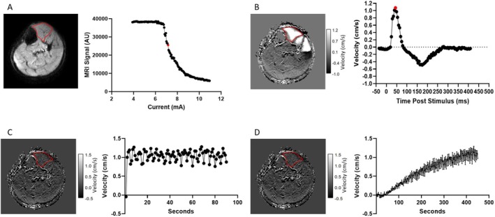
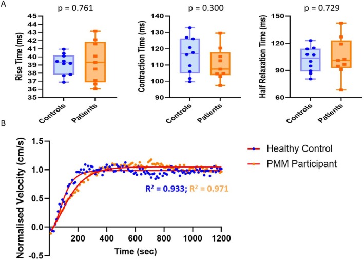
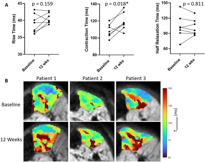
Muscle twitch dynamics and fatigability change in response to muscle disease. In this study, we developed an imaging paradigm to measure muscle twitch dynamics, and the response of the muscle to voluntary fatiguing contractions. We used a novel imaging technique called motor unit magnetic resonance imaging (MUMRI). MUMRI allows visualisation of muscle and motor unit activity by combining in-scanner electrical stimulation with dynamic pulsed gradient spin echo (twitch dynamics, PGSE-MUMRI) and phase contrast (fatigue, PC-MUMRI) imaging. In Part I of this study, we scanned 10 healthy controls, we measured the muscle rise (Trise), contraction (Tcontract) and half-relaxation time (Thalf-relax) of the tibialis anterior (TA) muscle on a voxel-wise basis using PGSE-MUMRI. Five controls were scanned twice to assess reproducibility; PGSE-MUMRI demonstrated reproducible results, with low variation between scans 3.4% for Trise, 6.4% for Tcontract and 7.1% for Thalf-relax. We then developed a PC-MUMRI paradigm to measure the recovery of the TA in response to a fatiguing voluntary exercise. In Part II of the study, we applied these two novel imaging paradigms in a cohort study of nine patients with single large-scale mtDNA deletion primary mitochondrial myopathy (PMM). Patients underwent a 12-week resistance exercise programme and baseline, and follow-up MRI was performed. PGSE-MUMRI detected a significantly longer muscle contraction time between baseline and follow-up in PMM patients 108.7 ± 7.9 vs. post-119.3 ± 10.4 ms; p = 0.018. There was no statistical difference in the recovery half maximum measured using PC-MUMRI in PMM patients between baseline and follow-up 254 ± 109 vs. 137 ± 41 s; p = 0.074. In conclusion, PGSE-MUMRI has detected differences in muscle twitch dynamics between controls and PMM following an exercise programme, and we can visualise differences in twitch dynamics subregions of muscle using this technique. The PC-MUMRI technique has shown promise as a novel measure of muscle fatigue.
期刊介绍:
NMR in Biomedicine is a journal devoted to the publication of original full-length papers, rapid communications and review articles describing the development of magnetic resonance spectroscopy or imaging methods or their use to investigate physiological, biochemical, biophysical or medical problems. Topics for submitted papers should be in one of the following general categories: (a) development of methods and instrumentation for MR of biological systems; (b) studies of normal or diseased organs, tissues or cells; (c) diagnosis or treatment of disease. Reports may cover work on patients or healthy human subjects, in vivo animal experiments, studies of isolated organs or cultured cells, analysis of tissue extracts, NMR theory, experimental techniques, or instrumentation.

 求助内容:
求助内容: 应助结果提醒方式:


