Mirjam Evi Braun, Francisco Fernandez Fernandez, Lena Riha, Hagen Schmal, Peter Schmittenbecher, Dorien Schneidmueller, Christoph Strüwind, Philipp Schwerk, Sebastian Reineke, Frank Traub, Christoph Ihle, Justus Lieber, Christina Wack, Hauke Rüther, Florian Baumann, Ingo Marzi, Lewin-Caspar Busse, Ludger Tüshaus, Miriam Adrian, Florian Bergmann, Alexander Graf, Martin M Kaiser, Oliver Loose
{"title":"Traumatic hip dislocations in children and adolescents: diagnostic challenges and the significance of MRI imaging-a multi-center study.","authors":"Mirjam Evi Braun, Francisco Fernandez Fernandez, Lena Riha, Hagen Schmal, Peter Schmittenbecher, Dorien Schneidmueller, Christoph Strüwind, Philipp Schwerk, Sebastian Reineke, Frank Traub, Christoph Ihle, Justus Lieber, Christina Wack, Hauke Rüther, Florian Baumann, Ingo Marzi, Lewin-Caspar Busse, Ludger Tüshaus, Miriam Adrian, Florian Bergmann, Alexander Graf, Martin M Kaiser, Oliver Loose","doi":"10.1007/s00068-025-02800-2","DOIUrl":null,"url":null,"abstract":"<p><strong>Background: </strong>Traumatic hip dislocations in children and adolescents are rare but can lead to severe outcomes like avascular necrosis. Delayed reductions, often due to overlooked dislocations in initial imaging, pose a major risk. The variability in symptoms and emergency care challenges early diagnosis. This multi-center study evaluates diagnostic approaches to enhance protocols for identifying traumatic hip dislocations in childhood.</p><p><strong>Methods: </strong>This retrospective multi-center study included 76 patients (aged ≤ 17 years) with acute traumatic hip dislocations and open growth plates from 16 German hospitals. Patient data and imaging from 1979 to 2022 were analyzed, with statistical evaluation performed using SPSS under ethical guidelines.</p><p><strong>Results: </strong>X-rays (single and biplanar views) were the primary diagnostic method, utilized in 85% of cases. Dislocations were missed in 12% (9 cases), primarily among children under eight years, with half of those under four. Delayed reductions (15.8%, n = 12) were linked to undetected dislocations in imaging in 9 cases. Conventional X-rays frequently missed dislocations, whereas MRI successfully identified all cases. Among the 76 patients, 54 (71%) had associated injuries, with 57.9% (n = 44) diagnosed exclusively via MRI.</p><p><strong>Conclusion: </strong>Timely diagnosis of traumatic hip dislocations is crucial, as delays increase the risk of femoral head necrosis. An algorithmic approach is essential for young children, where dislocations may not be readily suspected. MRI is vital in the secondary diagnostic phase, providing superior visualization of associated injuries, including acetabular avulsions and soft tissue interpositions highlighting the need for integration of MRI into a unified diagnostic algorithm for children suspected of such injuries.</p><p><strong>Level of evidence: </strong>IV.</p>","PeriodicalId":12064,"journal":{"name":"European Journal of Trauma and Emergency Surgery","volume":"51 1","pages":"194"},"PeriodicalIF":2.2000,"publicationDate":"2025-05-06","publicationTypes":"Journal Article","fieldsOfStudy":null,"isOpenAccess":false,"openAccessPdf":"https://www.ncbi.nlm.nih.gov/pmc/articles/PMC12055902/pdf/","citationCount":"0","resultStr":null,"platform":"Semanticscholar","paperid":null,"PeriodicalName":"European Journal of Trauma and Emergency Surgery","FirstCategoryId":"3","ListUrlMain":"https://doi.org/10.1007/s00068-025-02800-2","RegionNum":3,"RegionCategory":"医学","ArticlePicture":[],"TitleCN":null,"AbstractTextCN":null,"PMCID":null,"EPubDate":"","PubModel":"","JCR":"Q2","JCRName":"EMERGENCY MEDICINE","Score":null,"Total":0}
引用次数: 0
Abstract
Background: Traumatic hip dislocations in children and adolescents are rare but can lead to severe outcomes like avascular necrosis. Delayed reductions, often due to overlooked dislocations in initial imaging, pose a major risk. The variability in symptoms and emergency care challenges early diagnosis. This multi-center study evaluates diagnostic approaches to enhance protocols for identifying traumatic hip dislocations in childhood.
Methods: This retrospective multi-center study included 76 patients (aged ≤ 17 years) with acute traumatic hip dislocations and open growth plates from 16 German hospitals. Patient data and imaging from 1979 to 2022 were analyzed, with statistical evaluation performed using SPSS under ethical guidelines.
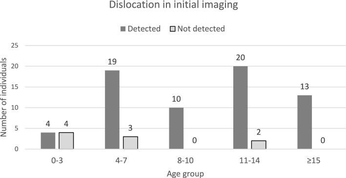
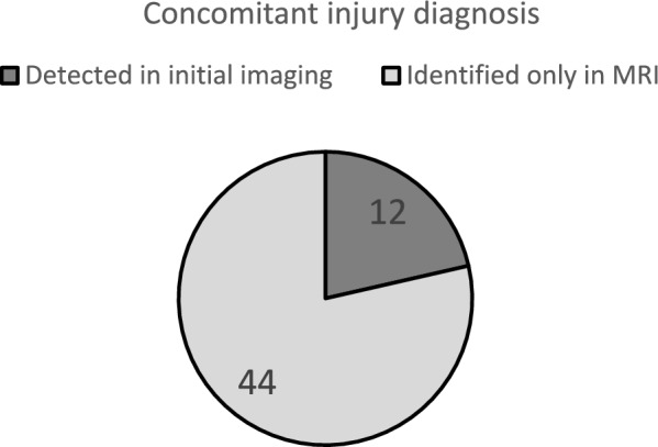
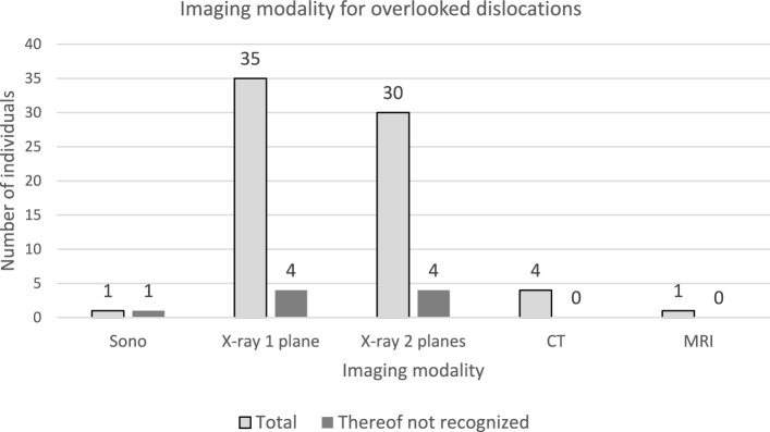
Results: X-rays (single and biplanar views) were the primary diagnostic method, utilized in 85% of cases. Dislocations were missed in 12% (9 cases), primarily among children under eight years, with half of those under four. Delayed reductions (15.8%, n = 12) were linked to undetected dislocations in imaging in 9 cases. Conventional X-rays frequently missed dislocations, whereas MRI successfully identified all cases. Among the 76 patients, 54 (71%) had associated injuries, with 57.9% (n = 44) diagnosed exclusively via MRI.
Conclusion: Timely diagnosis of traumatic hip dislocations is crucial, as delays increase the risk of femoral head necrosis. An algorithmic approach is essential for young children, where dislocations may not be readily suspected. MRI is vital in the secondary diagnostic phase, providing superior visualization of associated injuries, including acetabular avulsions and soft tissue interpositions highlighting the need for integration of MRI into a unified diagnostic algorithm for children suspected of such injuries.
期刊介绍:
The European Journal of Trauma and Emergency Surgery aims to open an interdisciplinary forum that allows for the scientific exchange between basic and clinical science related to pathophysiology, diagnostics and treatment of traumatized patients. The journal covers all aspects of clinical management, operative treatment and related research of traumatic injuries.
Clinical and experimental papers on issues relevant for the improvement of trauma care are published. Reviews, original articles, short communications and letters allow the appropriate presentation of major and minor topics.
 求助内容:
求助内容: 应助结果提醒方式:


