Christopher Keen, Justin Grenier, Peter Šereš, Robert Stobbe, James White, Christian Beaulieu, Rachel Sherrington, Amy Kirkham, D Ian Paterson, Richard Thompson
{"title":"MRI Assessment of Lung Water Density in Individuals Previously Infected With COVID-19: A Cross-Sectional Study.","authors":"Christopher Keen, Justin Grenier, Peter Šereš, Robert Stobbe, James White, Christian Beaulieu, Rachel Sherrington, Amy Kirkham, D Ian Paterson, Richard Thompson","doi":"10.1002/jmri.29814","DOIUrl":null,"url":null,"abstract":"<p><strong>Background: </strong>Lung damage in post-acute COVID-19 is a common clinical finding. Lung water density (LWD) imaging using ultrashort echo time (UTE) MRI with proton-density weighting is sensitive to edema and fibrosis.</p><p><strong>Purpose: </strong>To characterize LWD in COVID-19 survivors, compared with a healthy cohort.</p><p><strong>Study type: </strong>Retrospective cohort.</p><p><strong>Populations: </strong>185 COVID-19 survivors (63 male; age [median (interquartile range, IQR)]: 51 (25-83) years; 160 (66-363) days from COVID-19 infection to MRI) and 109 healthy controls (64 male; age: 52 (27-76) years) with no history of COVID-19 infection.</p><p><strong>Field strength/sequence: </strong>2.89T; Yarnball UTE pulse sequence.</p><p><strong>Assessment: </strong>Free-breathing three-dimensional LWD images were acquired in both cohorts. Clinical demographics (age, sex, body mass index [BMI]), presence of comorbidities (hypertension, dyslipidemia, diabetes, obesity), COVID-19 hospitalization, pulmonary function, six-minute walking distance, and plasma biomarkers were recorded.</p><p><strong>Statistical tests: </strong>Student's t-tests or Mann-Whitney U tests were used to compare lung water metrics between cohorts. The effect of comorbidities was assessed using Kruskal-Wallis tests followed by pairwise Wilcoxon tests with Bonferroni correction. Categorical variables were compared using chi-squared tests. p < 0.05 was considered significant.</p><p><strong>Results: </strong>LWD (median (IQR)), was significantly greater in the post-COVID-19 cohort than in the healthy cohort, 31.3 (6.6)% versus 27.9 (6.5)% in men and 30.3 (7.4)% versus 27.5 (4.9)% in women. 37% of men and 24% of women in the post-COVID-19 cohort had LWD above the healthy cohort 95% confidence limit. Participants with elevated LWD had significantly higher BMI (kg/m<sup>2</sup>) (32 (5) versus 26 (4) in men, 33 (9) versus 26 (7) in women), incidence of comorbidities (78% vs. 50% in men, 72% vs. 38% in women), rates of COVID-19 hospitalization (52% vs. 23% in men, 38% vs. 18% in women), and elevated CRP (mg/L) (2.2 (3.4) vs. 1.1 (1.4) in men, 1.8 (4.2) vs. 1.2 (2.1) in women).</p><p><strong>Data conclusion: </strong>MRI-derived LWD is elevated in COVID-19 survivors and is related to high BMI, COVID-19 hospitalization, inflammatory plasma biomarkers, and the presence of comorbidities.</p><p><strong>Evidence level: </strong>2.</p><p><strong>Technical efficacy: </strong>Stage 3.</p>","PeriodicalId":16140,"journal":{"name":"Journal of Magnetic Resonance Imaging","volume":" ","pages":"767-778"},"PeriodicalIF":3.5000,"publicationDate":"2025-09-01","publicationTypes":"Journal Article","fieldsOfStudy":null,"isOpenAccess":false,"openAccessPdf":"https://www.ncbi.nlm.nih.gov/pmc/articles/PMC12335338/pdf/","citationCount":"0","resultStr":null,"platform":"Semanticscholar","paperid":null,"PeriodicalName":"Journal of Magnetic Resonance Imaging","FirstCategoryId":"3","ListUrlMain":"https://doi.org/10.1002/jmri.29814","RegionNum":2,"RegionCategory":"医学","ArticlePicture":[],"TitleCN":null,"AbstractTextCN":null,"PMCID":null,"EPubDate":"2025/5/8 0:00:00","PubModel":"Epub","JCR":"Q1","JCRName":"RADIOLOGY, NUCLEAR MEDICINE & MEDICAL IMAGING","Score":null,"Total":0}
引用次数: 0
Abstract
Background: Lung damage in post-acute COVID-19 is a common clinical finding. Lung water density (LWD) imaging using ultrashort echo time (UTE) MRI with proton-density weighting is sensitive to edema and fibrosis.
Purpose: To characterize LWD in COVID-19 survivors, compared with a healthy cohort.
Study type: Retrospective cohort.
Populations: 185 COVID-19 survivors (63 male; age [median (interquartile range, IQR)]: 51 (25-83) years; 160 (66-363) days from COVID-19 infection to MRI) and 109 healthy controls (64 male; age: 52 (27-76) years) with no history of COVID-19 infection.
Field strength/sequence: 2.89T; Yarnball UTE pulse sequence.
Assessment: Free-breathing three-dimensional LWD images were acquired in both cohorts. Clinical demographics (age, sex, body mass index [BMI]), presence of comorbidities (hypertension, dyslipidemia, diabetes, obesity), COVID-19 hospitalization, pulmonary function, six-minute walking distance, and plasma biomarkers were recorded.
Statistical tests: Student's t-tests or Mann-Whitney U tests were used to compare lung water metrics between cohorts. The effect of comorbidities was assessed using Kruskal-Wallis tests followed by pairwise Wilcoxon tests with Bonferroni correction. Categorical variables were compared using chi-squared tests. p < 0.05 was considered significant.
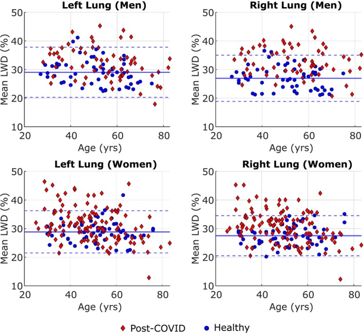
Results: LWD (median (IQR)), was significantly greater in the post-COVID-19 cohort than in the healthy cohort, 31.3 (6.6)% versus 27.9 (6.5)% in men and 30.3 (7.4)% versus 27.5 (4.9)% in women. 37% of men and 24% of women in the post-COVID-19 cohort had LWD above the healthy cohort 95% confidence limit. Participants with elevated LWD had significantly higher BMI (kg/m2) (32 (5) versus 26 (4) in men, 33 (9) versus 26 (7) in women), incidence of comorbidities (78% vs. 50% in men, 72% vs. 38% in women), rates of COVID-19 hospitalization (52% vs. 23% in men, 38% vs. 18% in women), and elevated CRP (mg/L) (2.2 (3.4) vs. 1.1 (1.4) in men, 1.8 (4.2) vs. 1.2 (2.1) in women).
Data conclusion: MRI-derived LWD is elevated in COVID-19 survivors and is related to high BMI, COVID-19 hospitalization, inflammatory plasma biomarkers, and the presence of comorbidities.
期刊介绍:
The Journal of Magnetic Resonance Imaging (JMRI) is an international journal devoted to the timely publication of basic and clinical research, educational and review articles, and other information related to the diagnostic applications of magnetic resonance.

 求助内容:
求助内容: 应助结果提醒方式:


