Jooyeon Lee, Jeong-Ran Park, Hanbyeol Lee, Seok-Ho Hong, Woo Jin Kim, Oliver Eickelberg, Sung-Min Park, Semin Ryu, Sung Joon Cho, Seung-Jin Kim, Se-Ran Yang
{"title":"Fludarabine attenuates inflammation and dysregulated autophagy in alveolar macrophages via inhibition of STAT1/IRF1 pathway.","authors":"Jooyeon Lee, Jeong-Ran Park, Hanbyeol Lee, Seok-Ho Hong, Woo Jin Kim, Oliver Eickelberg, Sung-Min Park, Semin Ryu, Sung Joon Cho, Seung-Jin Kim, Se-Ran Yang","doi":"10.1186/s42826-025-00245-7","DOIUrl":null,"url":null,"abstract":"<p><strong>Background: </strong>Acute lung injury (ALI), including its most severe form, acute respiratory distress syndrome (ARDS), is a common cause of acute hypoxemic respiratory failure. Although its clinical characteristics have been well characterized, the relevant mechanism remains unclear. An imbalance in autophagy leads to alveolar remodeling and triggers the pathogenesis of ARDS. In this study, we assessed the therapeutic efficacy of the STAT1 inhibitor fludarabine (Fluda) in ALI. C57BL6 mice were exposed to lipopolysaccharide (LPS), and their lung tissues were analyzed via next-generation transcriptome sequencing.</p><p><strong>Results: </strong>Western blotting revealed that interferon regulatory factor 1 (IRF1) was highly expressed and STAT1 was phosphorylated following LPS exposure. Fluda significantly decreased the protein expression of STAT1/IRF1 and inhibited the alveolar infiltration of neutrophils and macrophages. Nitric oxide (NO), inducible nitric oxide synthase, tumor necrosis factor-α (TNF-α), interferon-γ, and interleukin-6 (IL-6) release was decreased in the lungs of mice and RAW264.7 macrophages following Fluda treatment. In LPS-induced GFP-LC3 transgenic mice treated with Fluda, the counts of LC3-expressing neutrophils and macrophages in bronchoalveolar (BAL) fluid were significantly decreased. Furthermore, Fluda decreased LC3 and p62 protein expression, thereby inhibiting the release of NO, IL-6, and TNF-α in BAL. In RAW264.7 cells, the inhibition of STAT1/IRF1 by Fluda decreased LPS-induced ERK and NF-κB p65 phosphorylation.</p><p><strong>Conclusions: </strong>The inhibition of STAT1/IRF1 by Fluda plays a pivotal role in modulating dysregulated autophagy by suppressing the MAPK and NF-κB p65 pathways in ALI.</p>","PeriodicalId":17993,"journal":{"name":"Laboratory Animal Research","volume":"41 1","pages":"12"},"PeriodicalIF":2.9000,"publicationDate":"2025-05-07","publicationTypes":"Journal Article","fieldsOfStudy":null,"isOpenAccess":false,"openAccessPdf":"https://www.ncbi.nlm.nih.gov/pmc/articles/PMC12057031/pdf/","citationCount":"0","resultStr":null,"platform":"Semanticscholar","paperid":null,"PeriodicalName":"Laboratory Animal Research","FirstCategoryId":"1085","ListUrlMain":"https://doi.org/10.1186/s42826-025-00245-7","RegionNum":0,"RegionCategory":null,"ArticlePicture":[],"TitleCN":null,"AbstractTextCN":null,"PMCID":null,"EPubDate":"","PubModel":"","JCR":"Q3","JCRName":"MEDICINE, RESEARCH & EXPERIMENTAL","Score":null,"Total":0}
引用次数: 0
Abstract
Background: Acute lung injury (ALI), including its most severe form, acute respiratory distress syndrome (ARDS), is a common cause of acute hypoxemic respiratory failure. Although its clinical characteristics have been well characterized, the relevant mechanism remains unclear. An imbalance in autophagy leads to alveolar remodeling and triggers the pathogenesis of ARDS. In this study, we assessed the therapeutic efficacy of the STAT1 inhibitor fludarabine (Fluda) in ALI. C57BL6 mice were exposed to lipopolysaccharide (LPS), and their lung tissues were analyzed via next-generation transcriptome sequencing.
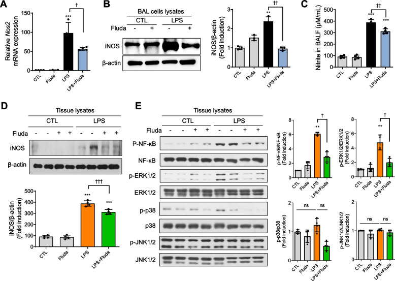
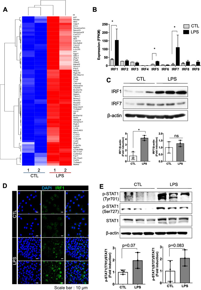
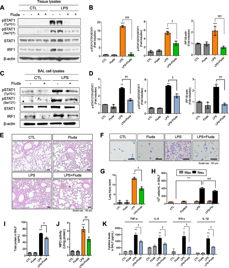
Results: Western blotting revealed that interferon regulatory factor 1 (IRF1) was highly expressed and STAT1 was phosphorylated following LPS exposure. Fluda significantly decreased the protein expression of STAT1/IRF1 and inhibited the alveolar infiltration of neutrophils and macrophages. Nitric oxide (NO), inducible nitric oxide synthase, tumor necrosis factor-α (TNF-α), interferon-γ, and interleukin-6 (IL-6) release was decreased in the lungs of mice and RAW264.7 macrophages following Fluda treatment. In LPS-induced GFP-LC3 transgenic mice treated with Fluda, the counts of LC3-expressing neutrophils and macrophages in bronchoalveolar (BAL) fluid were significantly decreased. Furthermore, Fluda decreased LC3 and p62 protein expression, thereby inhibiting the release of NO, IL-6, and TNF-α in BAL. In RAW264.7 cells, the inhibition of STAT1/IRF1 by Fluda decreased LPS-induced ERK and NF-κB p65 phosphorylation.
Conclusions: The inhibition of STAT1/IRF1 by Fluda plays a pivotal role in modulating dysregulated autophagy by suppressing the MAPK and NF-κB p65 pathways in ALI.

 求助内容:
求助内容: 应助结果提醒方式:


