Ashley Y Gao, Ameay V Naravane, Michael A Simmons, Tyler Looysen, Sandra Montezuma, Dara Koozekanani, Hossein Nazari
{"title":"Fulminant endophthalmitis after open globe injury by cat claw: two case reports and literature review.","authors":"Ashley Y Gao, Ameay V Naravane, Michael A Simmons, Tyler Looysen, Sandra Montezuma, Dara Koozekanani, Hossein Nazari","doi":"10.1186/s12348-025-00487-5","DOIUrl":null,"url":null,"abstract":"<p><strong>Objective: </strong>To describe two patients with fulminant endophthalmitis after penetrating ocular injuries by cat claw and review the literature regarding animal-related endophthalmitis.</p><p><strong>Design: </strong>Case series.</p><p><strong>Participants: </strong>In the study period, 298 patients were identified with a diagnosis of endophthalmitis, of which two were identified in association with open globe injury by cat claw.</p><p><strong>Methods: </strong>All patients with endophthalmitis after cat claw open globe injury in an academic center in a 20-year period are reported. Clinical and laboratory presentations, medical and surgical treatment, and outcomes are described. A literature review is summarized.</p><p><strong>Exposure: </strong>Open globe injury by cat claw.</p><p><strong>Main outcome measures: </strong>Interventions and ocular anatomical and functional outcomes.</p><p><strong>Results: </strong>Case 1: A 27-year-old female sustained a penetrating injury of the left eye by a cat claw. The laceration was repaired the next day, and intravitreal antibiotics injections were given. She developed acute fulminant endophthalmitis the following day and underwent pars plana vitrectomy, anterior chamber washout, and intravitreal antibiotics injection. Cultures isolated Propionibacterium acnes. A retinal detachment was noted after 48 days, requiring a second pars plana vitrectomy and tamponade with sulfur hexafluoride gas. The retina remained attached. Visual acuity at 14 months follow-up was 20/200. Case 2: A 42-year-old male developed endophthalmitis two days after a penetrating injury of the right eye by a cat claw. Pars plana vitrectomy and intravitreal antibiotics injections were performed the same day. Cultures identified Pasteurella multocida. The patient progressed to panophthalmitis in 24 h and received intravenous antibiotics. He developed proliferative vitreoretinopathy with recurrent retinal detachments requiring multiple vitrectomies. His visual acuity was hand motions at 7 months follow-up.</p><p><strong>Conclusions and relevance: </strong>Open globe injuries caused by cat claw may result in hyperacute and acute endophthalmitis. Propionibacterium acnes and Pasteurella multocida were isolated from the two cases reported here. Despite immediate interventions, both patients developed retinal detachment and had poor final visual acuity. Our report reveals that endophthalmitis caused by animal trauma is rare with potentially devastating outcomes, thereby requiring timely diagnosis and treatment.</p>","PeriodicalId":16600,"journal":{"name":"Journal of Ophthalmic Inflammation and Infection","volume":"15 1","pages":"41"},"PeriodicalIF":2.3000,"publicationDate":"2025-05-06","publicationTypes":"Journal Article","fieldsOfStudy":null,"isOpenAccess":false,"openAccessPdf":"https://www.ncbi.nlm.nih.gov/pmc/articles/PMC12055709/pdf/","citationCount":"0","resultStr":null,"platform":"Semanticscholar","paperid":null,"PeriodicalName":"Journal of Ophthalmic Inflammation and Infection","FirstCategoryId":"1085","ListUrlMain":"https://doi.org/10.1186/s12348-025-00487-5","RegionNum":0,"RegionCategory":null,"ArticlePicture":[],"TitleCN":null,"AbstractTextCN":null,"PMCID":null,"EPubDate":"","PubModel":"","JCR":"Q1","JCRName":"OPHTHALMOLOGY","Score":null,"Total":0}
引用次数: 0
Abstract
Objective: To describe two patients with fulminant endophthalmitis after penetrating ocular injuries by cat claw and review the literature regarding animal-related endophthalmitis.
Design: Case series.
Participants: In the study period, 298 patients were identified with a diagnosis of endophthalmitis, of which two were identified in association with open globe injury by cat claw.
Methods: All patients with endophthalmitis after cat claw open globe injury in an academic center in a 20-year period are reported. Clinical and laboratory presentations, medical and surgical treatment, and outcomes are described. A literature review is summarized.
Exposure: Open globe injury by cat claw.
Main outcome measures: Interventions and ocular anatomical and functional outcomes.
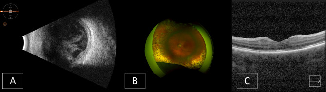
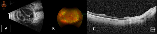
Results: Case 1: A 27-year-old female sustained a penetrating injury of the left eye by a cat claw. The laceration was repaired the next day, and intravitreal antibiotics injections were given. She developed acute fulminant endophthalmitis the following day and underwent pars plana vitrectomy, anterior chamber washout, and intravitreal antibiotics injection. Cultures isolated Propionibacterium acnes. A retinal detachment was noted after 48 days, requiring a second pars plana vitrectomy and tamponade with sulfur hexafluoride gas. The retina remained attached. Visual acuity at 14 months follow-up was 20/200. Case 2: A 42-year-old male developed endophthalmitis two days after a penetrating injury of the right eye by a cat claw. Pars plana vitrectomy and intravitreal antibiotics injections were performed the same day. Cultures identified Pasteurella multocida. The patient progressed to panophthalmitis in 24 h and received intravenous antibiotics. He developed proliferative vitreoretinopathy with recurrent retinal detachments requiring multiple vitrectomies. His visual acuity was hand motions at 7 months follow-up.
Conclusions and relevance: Open globe injuries caused by cat claw may result in hyperacute and acute endophthalmitis. Propionibacterium acnes and Pasteurella multocida were isolated from the two cases reported here. Despite immediate interventions, both patients developed retinal detachment and had poor final visual acuity. Our report reveals that endophthalmitis caused by animal trauma is rare with potentially devastating outcomes, thereby requiring timely diagnosis and treatment.
 求助内容:
求助内容: 应助结果提醒方式:


