Kamran Rakhshan, Ali Mohammadkhanizadeh, Mahdi Saberi Pirouz, Yaser Azizi
{"title":"Diosgenin Ameliorates Cardiac Function following Myocardial Ischemia Through Angiogenic and Anti-Fibrotic Properties; An Experimental Study.","authors":"Kamran Rakhshan, Ali Mohammadkhanizadeh, Mahdi Saberi Pirouz, Yaser Azizi","doi":"10.22037/aaemj.v13i1.2483","DOIUrl":null,"url":null,"abstract":"<p><strong>Introduction: </strong>Angiogenesis through restoration of blood supply to the ischemic myocardium is a pivotal process that contributes to cardiac repair and leads to improvement of myocardial function. This study was conducted to evaluate cardioprotective effects of Diosgenin against myocardial infarction (MI) with focus on angiogenesis, myocardial fibrosis, and oxidative stress.</p><p><strong>Methods: </strong>4 groups of male Wistar rats were considered for this study: (1) sham, (2) MI, (3) MI+Vehicle and (4) MI+Diosgenin. MI model was created by occluding left anterior descending (LAD) artery for 30 minutes and reperfusion was established for 14 days by opening this artery. Diosgenin (50 mg/kg) was given orally to the rats for 21 days (from 7 days before MI induction until the end of the 14-day reperfusion period). Cardiac injury markers including troponin I, creatine kinase-MB (CK-MB), and lactate dehydrogenase (LDH) were measured using enzyme-linked immunosorbent assay (ELISA), same as cardiac stress oxidative markers (superoxide dismutase (SOD), Malondialdehyde (MDA), reduced glutathione (GSH)). Echocardiography was used to measure heart function parameters and myocardial fibrosis was assessed via a specific tissue staining named Masson׳s trichrome. Blood vessel staining kit was used to assess left ventricular angiogenesis.</p><p><strong>Results: </strong>Ischemia-reperfusion injury increased serum levels of troponin I, CK-MB and LDH, as well as cardiac malondialdehyde (MDA) and myocardial fibrosis. MI also decreased myocardial function (Ejection fraction (EF)% and Fractional shortening (FS)%) and Diosgenin treatment reversed these parameters. Capillary density as marker of angiogenesis significantly increased in all of MI groups. However, development of angiogenesis was significantly higher in Diosgenin group compared with MI group.</p><p><strong>Conclusion: </strong>Diosgenin exerts cardioprotective effects against ischemia-reperfusion injury by strengthening cardiac antioxidant defense and reducing deposition of collagen fibers. It seems that the strengthening of angiogenesis in heart tissue is one of the main mechanisms of Diosgenin to increase the heart's resistance against ischemia.</p>","PeriodicalId":8146,"journal":{"name":"Archives of Academic Emergency Medicine","volume":"13 1","pages":"e40"},"PeriodicalIF":2.0000,"publicationDate":"2025-03-17","publicationTypes":"Journal Article","fieldsOfStudy":null,"isOpenAccess":false,"openAccessPdf":"https://www.ncbi.nlm.nih.gov/pmc/articles/PMC12065035/pdf/","citationCount":"0","resultStr":null,"platform":"Semanticscholar","paperid":null,"PeriodicalName":"Archives of Academic Emergency Medicine","FirstCategoryId":"1085","ListUrlMain":"https://doi.org/10.22037/aaemj.v13i1.2483","RegionNum":0,"RegionCategory":null,"ArticlePicture":[],"TitleCN":null,"AbstractTextCN":null,"PMCID":null,"EPubDate":"2025/1/1 0:00:00","PubModel":"eCollection","JCR":"Q1","JCRName":"EMERGENCY MEDICINE","Score":null,"Total":0}
引用次数: 0
Abstract
Introduction: Angiogenesis through restoration of blood supply to the ischemic myocardium is a pivotal process that contributes to cardiac repair and leads to improvement of myocardial function. This study was conducted to evaluate cardioprotective effects of Diosgenin against myocardial infarction (MI) with focus on angiogenesis, myocardial fibrosis, and oxidative stress.
Methods: 4 groups of male Wistar rats were considered for this study: (1) sham, (2) MI, (3) MI+Vehicle and (4) MI+Diosgenin. MI model was created by occluding left anterior descending (LAD) artery for 30 minutes and reperfusion was established for 14 days by opening this artery. Diosgenin (50 mg/kg) was given orally to the rats for 21 days (from 7 days before MI induction until the end of the 14-day reperfusion period). Cardiac injury markers including troponin I, creatine kinase-MB (CK-MB), and lactate dehydrogenase (LDH) were measured using enzyme-linked immunosorbent assay (ELISA), same as cardiac stress oxidative markers (superoxide dismutase (SOD), Malondialdehyde (MDA), reduced glutathione (GSH)). Echocardiography was used to measure heart function parameters and myocardial fibrosis was assessed via a specific tissue staining named Masson׳s trichrome. Blood vessel staining kit was used to assess left ventricular angiogenesis.
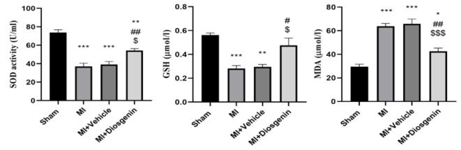
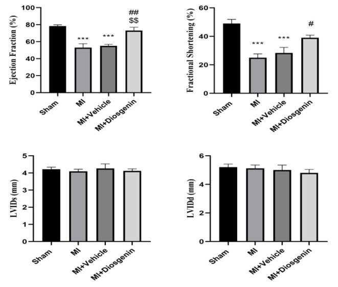
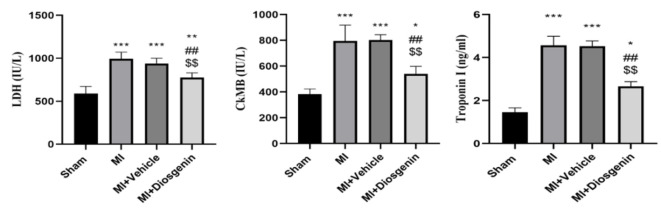
Results: Ischemia-reperfusion injury increased serum levels of troponin I, CK-MB and LDH, as well as cardiac malondialdehyde (MDA) and myocardial fibrosis. MI also decreased myocardial function (Ejection fraction (EF)% and Fractional shortening (FS)%) and Diosgenin treatment reversed these parameters. Capillary density as marker of angiogenesis significantly increased in all of MI groups. However, development of angiogenesis was significantly higher in Diosgenin group compared with MI group.
Conclusion: Diosgenin exerts cardioprotective effects against ischemia-reperfusion injury by strengthening cardiac antioxidant defense and reducing deposition of collagen fibers. It seems that the strengthening of angiogenesis in heart tissue is one of the main mechanisms of Diosgenin to increase the heart's resistance against ischemia.

 求助内容:
求助内容: 应助结果提醒方式:


