A Teh, R Crisman, E Dwars, R Malik, L H de Miranda, W Meyer, M Krockenberger
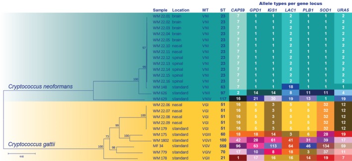
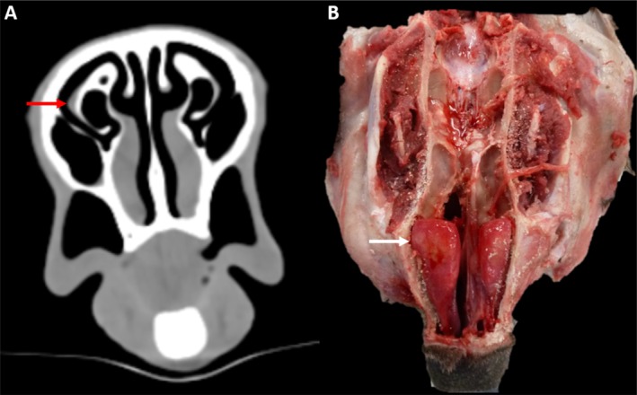
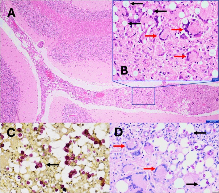
{"title":"Cryptococcal meningitis due to Cryptococcus neoformans VNI in a koala (Phascolarctos cinereus) with progressive neurological disease.","authors":"A Teh, R Crisman, E Dwars, R Malik, L H de Miranda, W Meyer, M Krockenberger","doi":"10.1111/avj.13446","DOIUrl":null,"url":null,"abstract":"<p><p>Nearly all cases of cryptococcosis in koalas are caused by Cryptococcus gattii species complex. A rare case of meningitis due to Cryptococcus neoformans VNI/AFLP1 (abbreviated VNI) is described in a koala with nasal colonisation by both species complexes. An eight-year-old koala in a wildlife park presented for seizures and returned a positive latex cryptococcal antigen agglutination test. The koala was euthanased due to the severity of disease, and a post-mortem computed tomography study showed mild mucosal thickening of the right nasal turbinates. The necropsy also showed slightly turbid cisternal cerebrospinal fluid and meningeal opacity. Histology revealed severe granulomatous cryptococcal meningitis and paucireactive right cryptococcal rhinitis. Fungal cultures yielded heavy pure growths of C. neoformans from the brain and spinal cord, and comparable heavy growths of both C. neoformans and C. gattii from the nasal cavity. Cryptococcus species complexes were identified by mass spectrometry (MALDI-TOF) and multi-locus sequence typing (MLST). The C. neoformans isolates from the brain, spinal cord and nasal cavity were identical by MLST and classified as sequencing type (ST) 23 and molecular type (MT) VNI. The C. gattii isolates were classified as ST 51 and AFLP4/VGI (abbreviated VGI). This suggests that the meningitis developed as an extension of C. neoformans VNI from nasal cavity colonisation. This is the second documented case of central nervous system (CNS) cryptococcosis due to C. neoformans species complex in a koala and the first in Australia. Despite heavy nasal colonisation by C. gattii, only the C. neoformans isolate progressed to meningitis.</p>","PeriodicalId":8661,"journal":{"name":"Australian Veterinary Journal","volume":" ","pages":"487-493"},"PeriodicalIF":1.7000,"publicationDate":"2025-08-01","publicationTypes":"Journal Article","fieldsOfStudy":null,"isOpenAccess":false,"openAccessPdf":"https://www.ncbi.nlm.nih.gov/pmc/articles/PMC12331396/pdf/","citationCount":"0","resultStr":null,"platform":"Semanticscholar","paperid":null,"PeriodicalName":"Australian Veterinary Journal","FirstCategoryId":"97","ListUrlMain":"https://doi.org/10.1111/avj.13446","RegionNum":4,"RegionCategory":"农林科学","ArticlePicture":[],"TitleCN":null,"AbstractTextCN":null,"PMCID":null,"EPubDate":"2025/4/22 0:00:00","PubModel":"Epub","JCR":"Q2","JCRName":"VETERINARY SCIENCES","Score":null,"Total":0}
引用次数: 0
Abstract
Nearly all cases of cryptococcosis in koalas are caused by Cryptococcus gattii species complex. A rare case of meningitis due to Cryptococcus neoformans VNI/AFLP1 (abbreviated VNI) is described in a koala with nasal colonisation by both species complexes. An eight-year-old koala in a wildlife park presented for seizures and returned a positive latex cryptococcal antigen agglutination test. The koala was euthanased due to the severity of disease, and a post-mortem computed tomography study showed mild mucosal thickening of the right nasal turbinates. The necropsy also showed slightly turbid cisternal cerebrospinal fluid and meningeal opacity. Histology revealed severe granulomatous cryptococcal meningitis and paucireactive right cryptococcal rhinitis. Fungal cultures yielded heavy pure growths of C. neoformans from the brain and spinal cord, and comparable heavy growths of both C. neoformans and C. gattii from the nasal cavity. Cryptococcus species complexes were identified by mass spectrometry (MALDI-TOF) and multi-locus sequence typing (MLST). The C. neoformans isolates from the brain, spinal cord and nasal cavity were identical by MLST and classified as sequencing type (ST) 23 and molecular type (MT) VNI. The C. gattii isolates were classified as ST 51 and AFLP4/VGI (abbreviated VGI). This suggests that the meningitis developed as an extension of C. neoformans VNI from nasal cavity colonisation. This is the second documented case of central nervous system (CNS) cryptococcosis due to C. neoformans species complex in a koala and the first in Australia. Despite heavy nasal colonisation by C. gattii, only the C. neoformans isolate progressed to meningitis.
期刊介绍:
Over the past 80 years, the Australian Veterinary Journal (AVJ) has been providing the veterinary profession with leading edge clinical and scientific research, case reports, reviews. news and timely coverage of industry issues. AJV is Australia''s premier veterinary science text and is distributed monthly to over 5,500 Australian Veterinary Association members and subscribers.


 求助内容:
求助内容: 应助结果提醒方式:


