{"title":"Multiple pulmonary mucinous cystadenoma with Ovarian-like stroma: a case report.","authors":"Lei Wang, Xieraili Wumener, Wenting Huang","doi":"10.1186/s13000-025-01642-1","DOIUrl":null,"url":null,"abstract":"<p><strong>Background: </strong>Pulmonary mucinous cystadenoma with ovarian-like stroma has rarely been reported. To the best of our knowledge, only two prior cases have been reported to date.</p><p><strong>Case presentation: </strong>A 47-year-old female underwent an <sup>18</sup>F-fluorodeoxyglucose(FDG) positron emission tomography/computed tomography(PET/CT) scan, revealig multiple solid and cystic nodules with mild FDG uptake in both lungs. The tumor exhibited an adenoid or papillary structure, covered by monolayer mucous columnar epithelium and pseudostratified ciliated columnar epithelium, with abundant mesenchymal cells. Nuclei were oval, fusiform, or polygonal, resembling ovarian-like stroma, and showed mild nuclear atypia without atypical mitoses. Immunohistochemical analysis indicated: CK (epithelial cell +), P63 (epithelial basal cells +), CK7 (epithelial cell +), CK20 (-), TTF-1 (epithelial cell +), napsin A (partial epithelial cell +), α-inhibin (-), S100 (-), SMA (-), EMA (epithelial cell +), CEA (-), WT-1 (mesenchymal cells +), ER (mesenchymal cells +), P16 (partial mesenchymal cells +), CD10 (mesenchymal cells +), and Ki-67 (2% +).</p><p><strong>Conclusion: </strong>Pulmonary mucinous cystadenoma with ovarian-like stroma is rare, and its pathological nature and classification are not yet fully understood.</p>","PeriodicalId":11237,"journal":{"name":"Diagnostic Pathology","volume":"20 1","pages":"44"},"PeriodicalIF":2.3000,"publicationDate":"2025-04-16","publicationTypes":"Journal Article","fieldsOfStudy":null,"isOpenAccess":false,"openAccessPdf":"https://www.ncbi.nlm.nih.gov/pmc/articles/PMC12001701/pdf/","citationCount":"0","resultStr":null,"platform":"Semanticscholar","paperid":null,"PeriodicalName":"Diagnostic Pathology","FirstCategoryId":"3","ListUrlMain":"https://doi.org/10.1186/s13000-025-01642-1","RegionNum":3,"RegionCategory":"医学","ArticlePicture":[],"TitleCN":null,"AbstractTextCN":null,"PMCID":null,"EPubDate":"","PubModel":"","JCR":"Q2","JCRName":"PATHOLOGY","Score":null,"Total":0}
引用次数: 0
Abstract
Background: Pulmonary mucinous cystadenoma with ovarian-like stroma has rarely been reported. To the best of our knowledge, only two prior cases have been reported to date.
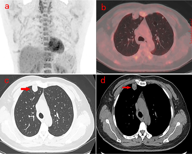
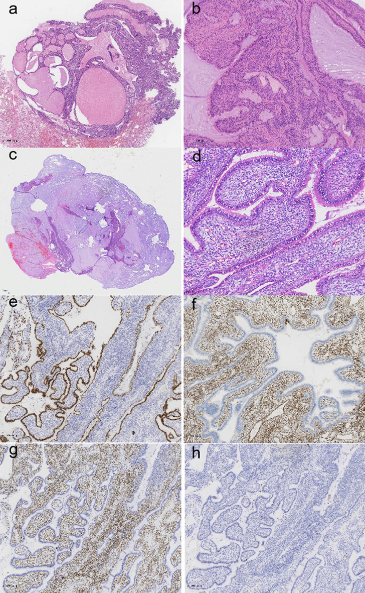
Case presentation: A 47-year-old female underwent an 18F-fluorodeoxyglucose(FDG) positron emission tomography/computed tomography(PET/CT) scan, revealig multiple solid and cystic nodules with mild FDG uptake in both lungs. The tumor exhibited an adenoid or papillary structure, covered by monolayer mucous columnar epithelium and pseudostratified ciliated columnar epithelium, with abundant mesenchymal cells. Nuclei were oval, fusiform, or polygonal, resembling ovarian-like stroma, and showed mild nuclear atypia without atypical mitoses. Immunohistochemical analysis indicated: CK (epithelial cell +), P63 (epithelial basal cells +), CK7 (epithelial cell +), CK20 (-), TTF-1 (epithelial cell +), napsin A (partial epithelial cell +), α-inhibin (-), S100 (-), SMA (-), EMA (epithelial cell +), CEA (-), WT-1 (mesenchymal cells +), ER (mesenchymal cells +), P16 (partial mesenchymal cells +), CD10 (mesenchymal cells +), and Ki-67 (2% +).
Conclusion: Pulmonary mucinous cystadenoma with ovarian-like stroma is rare, and its pathological nature and classification are not yet fully understood.
期刊介绍:
Diagnostic Pathology is an open access, peer-reviewed, online journal that considers research in surgical and clinical pathology, immunology, and biology, with a special focus on cutting-edge approaches in diagnostic pathology and tissue-based therapy. The journal covers all aspects of surgical pathology, including classic diagnostic pathology, prognosis-related diagnosis (tumor stages, prognosis markers, such as MIB-percentage, hormone receptors, etc.), and therapy-related findings. The journal also focuses on the technological aspects of pathology, including molecular biology techniques, morphometry aspects (stereology, DNA analysis, syntactic structure analysis), communication aspects (telecommunication, virtual microscopy, virtual pathology institutions, etc.), and electronic education and quality assurance (for example interactive publication, on-line references with automated updating, etc.).

 求助内容:
求助内容: 应助结果提醒方式:


