{"title":"Pulsed Nd:YAG laser therapy accelerates fracture healing in a rat femoral osteotomy model.","authors":"Po-Yen Ko, Che-Chia Hsu, Shih-Yao Chen, Chieh-Hsiang Hsu, Chia-Lung Li, I-Ming Jou, Po-Ting Wu","doi":"10.1302/2046-3758.145.BJR-2024-0285.R2","DOIUrl":null,"url":null,"abstract":"<p><strong>Aims: </strong>This study aimed to evaluate the effects of Nd:YAG laser treatment on fracture healing in a rat model. We hypothesized that laser therapy would accelerate healing by stimulating early neovascularization and osteoblast recruitment.</p><p><strong>Methods: </strong>A total of 54 male Sprague-Dawley rats received intramedullary Kirschner wire (K-wire) osteosynthesis following femoral osteotomy, and were randomly divided into two groups (n = 27 each): the control group, and the laser group that received daily pulsed Nd:YAG laser for ten days immediately after osteotomy. Fracture sites were assessed using micro-CT (μCT; n = 8 at each timepoint), histology (n = 4), and three-point bending tests (n = 4) at week 2, week 4<i>,</i> and week 6, respectively. At week 2, an additional three rats per group were selected for the western blot tests.</p><p><strong>Results: </strong>Compared to controls, the laser group showed higher vascular endothelial growth factor (VEGF), CD31, and Runx2 protein expression, and significantly higher neovascular area density and osteoblast density (p = 0.025 and p = 0.008, respectively) at week 2. At week 4, the laser treatment led to higher histological fracture healing scale and flexural modulus, and less strain (p = 0.001, p = 0.020, and p = 0.004, respectively). Macroscopically, the laser group showed higher mature bone volume fraction and radiological union score at weeks 4 and 6 (volume fraction: p = 0.017 and p = 0.001; union score: p = 0.001 and p = 0.024, respectively).</p><p><strong>Conclusion: </strong>Pulsed Nd:YAG laser therapy accelerates multiple quantitative indicators of fracture healing within six weeks in a rat femoral osteotomy model, which was associated with enhanced angiogenesis and osteogenesis during the early healing phase.</p>","PeriodicalId":9074,"journal":{"name":"Bone & Joint Research","volume":"14 5","pages":"376-388"},"PeriodicalIF":5.1000,"publicationDate":"2025-05-02","publicationTypes":"Journal Article","fieldsOfStudy":null,"isOpenAccess":false,"openAccessPdf":"https://www.ncbi.nlm.nih.gov/pmc/articles/PMC12045664/pdf/","citationCount":"0","resultStr":null,"platform":"Semanticscholar","paperid":null,"PeriodicalName":"Bone & Joint Research","FirstCategoryId":"3","ListUrlMain":"https://doi.org/10.1302/2046-3758.145.BJR-2024-0285.R2","RegionNum":2,"RegionCategory":"医学","ArticlePicture":[],"TitleCN":null,"AbstractTextCN":null,"PMCID":null,"EPubDate":"","PubModel":"","JCR":"Q2","JCRName":"CELL & TISSUE ENGINEERING","Score":null,"Total":0}
引用次数: 0
Abstract
Aims: This study aimed to evaluate the effects of Nd:YAG laser treatment on fracture healing in a rat model. We hypothesized that laser therapy would accelerate healing by stimulating early neovascularization and osteoblast recruitment.
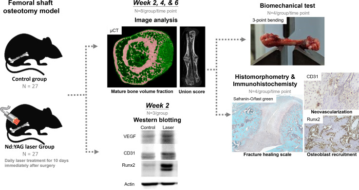
Methods: A total of 54 male Sprague-Dawley rats received intramedullary Kirschner wire (K-wire) osteosynthesis following femoral osteotomy, and were randomly divided into two groups (n = 27 each): the control group, and the laser group that received daily pulsed Nd:YAG laser for ten days immediately after osteotomy. Fracture sites were assessed using micro-CT (μCT; n = 8 at each timepoint), histology (n = 4), and three-point bending tests (n = 4) at week 2, week 4, and week 6, respectively. At week 2, an additional three rats per group were selected for the western blot tests.
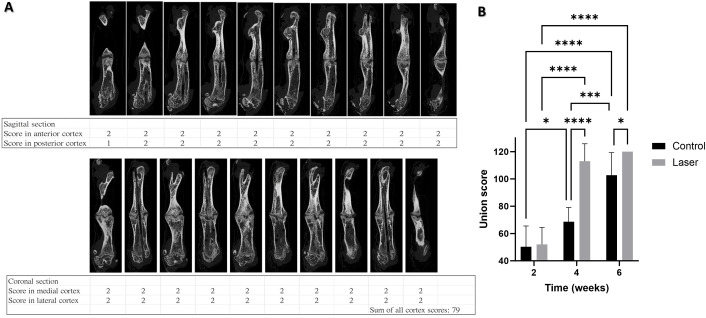
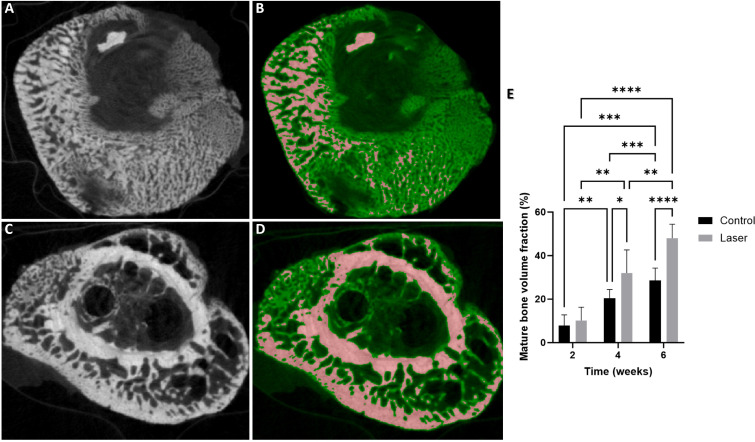
Results: Compared to controls, the laser group showed higher vascular endothelial growth factor (VEGF), CD31, and Runx2 protein expression, and significantly higher neovascular area density and osteoblast density (p = 0.025 and p = 0.008, respectively) at week 2. At week 4, the laser treatment led to higher histological fracture healing scale and flexural modulus, and less strain (p = 0.001, p = 0.020, and p = 0.004, respectively). Macroscopically, the laser group showed higher mature bone volume fraction and radiological union score at weeks 4 and 6 (volume fraction: p = 0.017 and p = 0.001; union score: p = 0.001 and p = 0.024, respectively).
Conclusion: Pulsed Nd:YAG laser therapy accelerates multiple quantitative indicators of fracture healing within six weeks in a rat femoral osteotomy model, which was associated with enhanced angiogenesis and osteogenesis during the early healing phase.
 求助内容:
求助内容: 应助结果提醒方式:


