Steven E. Johnson, Chad R. Haney, Alisha N. Spann, Nigar Khurram, Farres Obeidin, Jungwha Lee, Ming Zhao
{"title":"An in vivo imaging approach for simultaneously assessing tumor response and cytotoxicity-induced tissue response in chemotherapy","authors":"Steven E. Johnson, Chad R. Haney, Alisha N. Spann, Nigar Khurram, Farres Obeidin, Jungwha Lee, Ming Zhao","doi":"10.1007/s10495-025-02118-9","DOIUrl":null,"url":null,"abstract":"<div><p>In chemotherapeutic treatments, while cancer cells are the primary target, cytotoxic side effects are an important consideration. In the current study, we applied an in vivo imaging tool for characterizing chemotherapeutic response in a preclinical setting. The study focused on simultaneously examining the tumor and tissue response as a result of treatment with bortezomib, a mainstay proteasome inhibitor for treating multiple myeloma, in a preclinical model. OPM-2 tumor-bearing SCID-beige mice were designated as control or treated with bortezomib (1 mg/kg, i.v., every 4 days) (n = 8 per group). <sup>99m</sup>Tc-duramycin SPECT/CT whole-body scans were acquired 2 days before treatment as baseline and at days 1, 3 and 5 after treatment. Radioactivity uptake in tissues and organs was determined and quantitatively compared between control and bortezomib-treated group at each of the time points. Based on the imaging data, separate groups of tumor-bearing mice (n = 3 each) were included as control and bortezomib treated and the tissues were collected on day 5 for histopathology. In vivo imaging data identified significantly elevated <sup>99m</sup>Tc-duramycin uptake in the tumor, particularly in tumoral periphery. This was accompanied with signal changes in multiple organs and tissues including the adipose tissue, major bones, abdominal regions, spleen and testes. The imaging findings were consistent with known cytotoxic side effects of bortezomib and were supported by histopathology. The outcome of the study demonstrated potential utilities of the technology by enabling timely determination of the efficacy of anticancer treatments and the effect on collateral tissues as a result of systemic cytotoxic treatment.</p></div>","PeriodicalId":8062,"journal":{"name":"Apoptosis","volume":"30 5-6","pages":"1515 - 1524"},"PeriodicalIF":8.1000,"publicationDate":"2025-04-26","publicationTypes":"Journal Article","fieldsOfStudy":null,"isOpenAccess":false,"openAccessPdf":"https://www.ncbi.nlm.nih.gov/pmc/articles/PMC12167287/pdf/","citationCount":"0","resultStr":null,"platform":"Semanticscholar","paperid":null,"PeriodicalName":"Apoptosis","FirstCategoryId":"99","ListUrlMain":"https://link.springer.com/article/10.1007/s10495-025-02118-9","RegionNum":2,"RegionCategory":"生物学","ArticlePicture":[],"TitleCN":null,"AbstractTextCN":null,"PMCID":null,"EPubDate":"","PubModel":"","JCR":"Q1","JCRName":"BIOCHEMISTRY & MOLECULAR BIOLOGY","Score":null,"Total":0}
引用次数: 0
Abstract
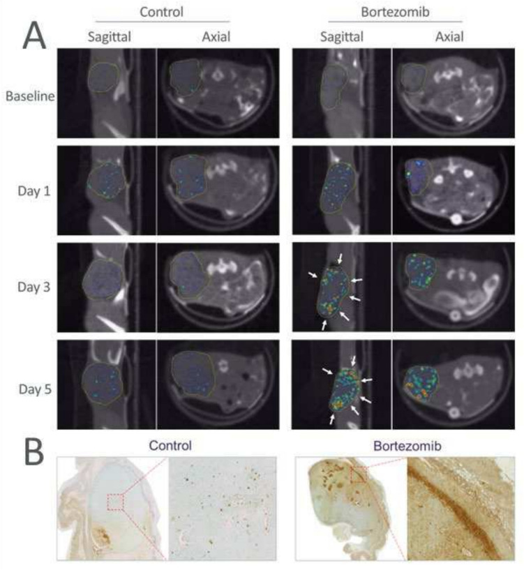
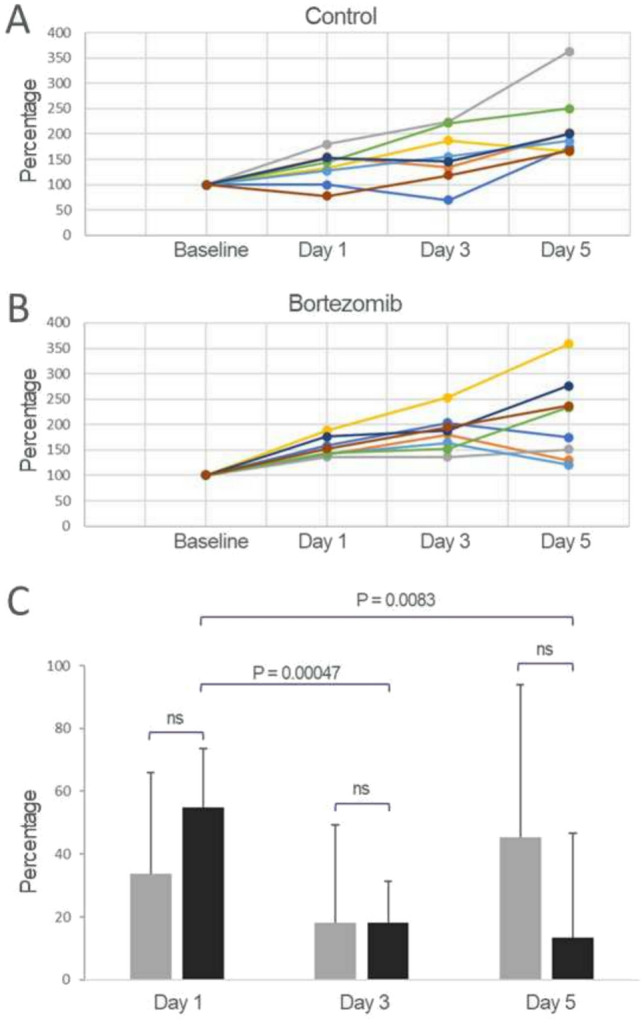
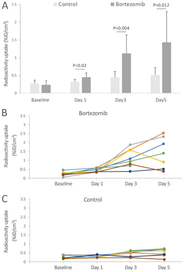
In chemotherapeutic treatments, while cancer cells are the primary target, cytotoxic side effects are an important consideration. In the current study, we applied an in vivo imaging tool for characterizing chemotherapeutic response in a preclinical setting. The study focused on simultaneously examining the tumor and tissue response as a result of treatment with bortezomib, a mainstay proteasome inhibitor for treating multiple myeloma, in a preclinical model. OPM-2 tumor-bearing SCID-beige mice were designated as control or treated with bortezomib (1 mg/kg, i.v., every 4 days) (n = 8 per group). 99mTc-duramycin SPECT/CT whole-body scans were acquired 2 days before treatment as baseline and at days 1, 3 and 5 after treatment. Radioactivity uptake in tissues and organs was determined and quantitatively compared between control and bortezomib-treated group at each of the time points. Based on the imaging data, separate groups of tumor-bearing mice (n = 3 each) were included as control and bortezomib treated and the tissues were collected on day 5 for histopathology. In vivo imaging data identified significantly elevated 99mTc-duramycin uptake in the tumor, particularly in tumoral periphery. This was accompanied with signal changes in multiple organs and tissues including the adipose tissue, major bones, abdominal regions, spleen and testes. The imaging findings were consistent with known cytotoxic side effects of bortezomib and were supported by histopathology. The outcome of the study demonstrated potential utilities of the technology by enabling timely determination of the efficacy of anticancer treatments and the effect on collateral tissues as a result of systemic cytotoxic treatment.
期刊介绍:
Apoptosis, a monthly international peer-reviewed journal, focuses on the rapid publication of innovative investigations into programmed cell death. The journal aims to stimulate research on the mechanisms and role of apoptosis in various human diseases, such as cancer, autoimmune disease, viral infection, AIDS, cardiovascular disease, neurodegenerative disorders, osteoporosis, and aging. The Editor-In-Chief acknowledges the importance of advancing clinical therapies for apoptosis-related diseases. Apoptosis considers Original Articles, Reviews, Short Communications, Letters to the Editor, and Book Reviews for publication.

 求助内容:
求助内容: 应助结果提醒方式:


