Lucas de Vries, M M Quirien Robbe, Ivo G H Jansen, S Mahsa Mojtahedi, Jan W Hoving, Susanne G H Olthuis, Robrecht R M M Knapen, Florentina M E Pinckaers, Manon Kappelhof, Ludo F M Beenen, Alida A Postma, Robert J van Oostenbrugge, Diederik W J Dippel, Efstratios Gavves, Bart J Emmer, Charles B L M Majoie, Wim H van Zwam, Henk A Marquering
{"title":"Automated collateral assessment restricted to the hypoperfused area for distal vessel occlusions in ischemic stroke.","authors":"Lucas de Vries, M M Quirien Robbe, Ivo G H Jansen, S Mahsa Mojtahedi, Jan W Hoving, Susanne G H Olthuis, Robrecht R M M Knapen, Florentina M E Pinckaers, Manon Kappelhof, Ludo F M Beenen, Alida A Postma, Robert J van Oostenbrugge, Diederik W J Dippel, Efstratios Gavves, Bart J Emmer, Charles B L M Majoie, Wim H van Zwam, Henk A Marquering","doi":"10.1007/s00330-025-11442-2","DOIUrl":null,"url":null,"abstract":"<p><strong>Objectives: </strong>This study aims to: (1) develop and evaluate a quantitative assessment of collateral status in the downstream area of an occluded intracranial artery in acute ischemic stroke and compare this method to middle cerebral artery (MCA)-based assessment; (2) determine the agreement between the automated occlusion-downstream area collateral score (ODACS) and expert raters' assessments, and compare this to inter-rater agreement.</p><p><strong>Methods: </strong>Patients from MR CLEAN-NO IV and MR CLEAN Registry with a proximal M1, distal M1, or M2 occlusion were included. Using the hypoperfused area from CT perfusion (CTP) as a proxy for the occlusion-downstream territory and automated vessel segmentations from CT angiography (CTA), ODACS is calculated as the vessel volume ratio between downstream ipsilateral and its contralateral regions. ODACS was compared to a whole MCA-territory approach and evaluated against visual scoring by two expert raters that visually estimated ODACS using CTA and CTP, and their inter-rater agreement.</p><p><strong>Results: </strong>The study included 204 patients with a proximal M1 (52%), distal M1 (32%), or M2 (16%) occlusion. ODACS yielded lower collateral scores than MCA-based scoring for all occlusion locations, with larger differences in more distal occlusions. For M2 occlusions, 58% of patients shifted from good (> 50%) to poor (≤ 50%) collateral filling of the occluded territory using ODACS. Moderate (weighted Cohen's kappa κ = 0.45) inter-rater agreement and fair (κ = 0.35) to moderate (κ = 0.51) ODACS-rater agreement were observed.</p><p><strong>Conclusions: </strong>ODACS yields lower collateral scores compared to MCA-based scoring and is comparable to scores from expert raters.</p><p><strong>Key points: </strong>Question CT angiography-based collateral assessment in the MCA territory is inadequate to assess the collateral status in patients with distal vessel occlusions. Findings Our automated ODACS revealed lower collateral scores than traditional whole-territory assessment, especially in distal vessel occlusions. Clinical relevance The more precise evaluation of affected brain territories through automated occlusion-downstream area assessments prevents an overestimation of collateral status in distal occlusions, which could lead to improved patient selection and treatment decisions in acute stroke care.</p>","PeriodicalId":12076,"journal":{"name":"European Radiology","volume":" ","pages":"6127-6139"},"PeriodicalIF":4.7000,"publicationDate":"2025-10-01","publicationTypes":"Journal Article","fieldsOfStudy":null,"isOpenAccess":false,"openAccessPdf":"https://www.ncbi.nlm.nih.gov/pmc/articles/PMC12417235/pdf/","citationCount":"0","resultStr":null,"platform":"Semanticscholar","paperid":null,"PeriodicalName":"European Radiology","FirstCategoryId":"3","ListUrlMain":"https://doi.org/10.1007/s00330-025-11442-2","RegionNum":2,"RegionCategory":"医学","ArticlePicture":[],"TitleCN":null,"AbstractTextCN":null,"PMCID":null,"EPubDate":"2025/4/14 0:00:00","PubModel":"Epub","JCR":"Q1","JCRName":"RADIOLOGY, NUCLEAR MEDICINE & MEDICAL IMAGING","Score":null,"Total":0}
引用次数: 0
Abstract
Objectives: This study aims to: (1) develop and evaluate a quantitative assessment of collateral status in the downstream area of an occluded intracranial artery in acute ischemic stroke and compare this method to middle cerebral artery (MCA)-based assessment; (2) determine the agreement between the automated occlusion-downstream area collateral score (ODACS) and expert raters' assessments, and compare this to inter-rater agreement.
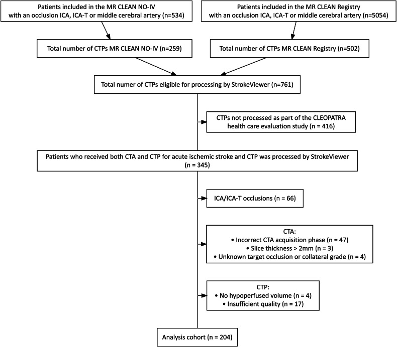
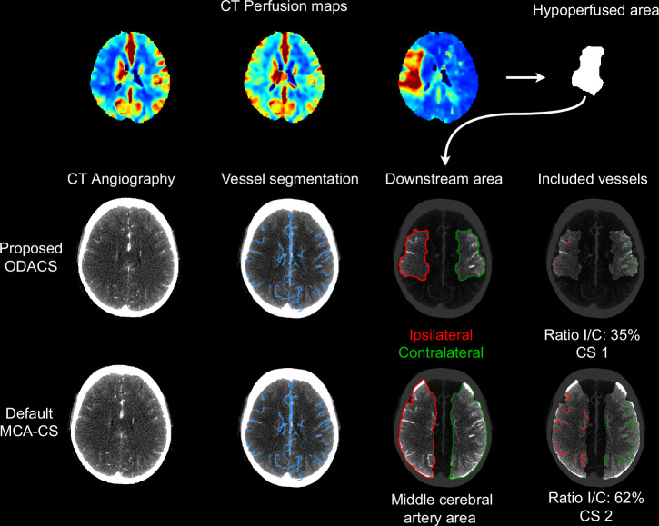
Methods: Patients from MR CLEAN-NO IV and MR CLEAN Registry with a proximal M1, distal M1, or M2 occlusion were included. Using the hypoperfused area from CT perfusion (CTP) as a proxy for the occlusion-downstream territory and automated vessel segmentations from CT angiography (CTA), ODACS is calculated as the vessel volume ratio between downstream ipsilateral and its contralateral regions. ODACS was compared to a whole MCA-territory approach and evaluated against visual scoring by two expert raters that visually estimated ODACS using CTA and CTP, and their inter-rater agreement.
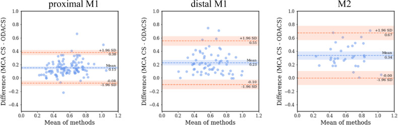
Results: The study included 204 patients with a proximal M1 (52%), distal M1 (32%), or M2 (16%) occlusion. ODACS yielded lower collateral scores than MCA-based scoring for all occlusion locations, with larger differences in more distal occlusions. For M2 occlusions, 58% of patients shifted from good (> 50%) to poor (≤ 50%) collateral filling of the occluded territory using ODACS. Moderate (weighted Cohen's kappa κ = 0.45) inter-rater agreement and fair (κ = 0.35) to moderate (κ = 0.51) ODACS-rater agreement were observed.
Conclusions: ODACS yields lower collateral scores compared to MCA-based scoring and is comparable to scores from expert raters.
Key points: Question CT angiography-based collateral assessment in the MCA territory is inadequate to assess the collateral status in patients with distal vessel occlusions. Findings Our automated ODACS revealed lower collateral scores than traditional whole-territory assessment, especially in distal vessel occlusions. Clinical relevance The more precise evaluation of affected brain territories through automated occlusion-downstream area assessments prevents an overestimation of collateral status in distal occlusions, which could lead to improved patient selection and treatment decisions in acute stroke care.
期刊介绍:
European Radiology (ER) continuously updates scientific knowledge in radiology by publication of strong original articles and state-of-the-art reviews written by leading radiologists. A well balanced combination of review articles, original papers, short communications from European radiological congresses and information on society matters makes ER an indispensable source for current information in this field.
This is the Journal of the European Society of Radiology, and the official journal of a number of societies.
From 2004-2008 supplements to European Radiology were published under its companion, European Radiology Supplements, ISSN 1613-3749.

 求助内容:
求助内容: 应助结果提醒方式:


