Vahid RoshanRavan, Hamidreza Ghorbani, Salman Soltani, Elham Shabani, Hosein Tanha, Ali Moradi, Kamran Aryana, Hasan Mehrad Majd, Behzad Aminzadeh, Reza Jafaei Daloei, Keyvan Sadri, Mahdi Momen Nejhad, Atena Aghaee
{"title":"The diagnostic value of whole-body HYNIC-PSMA 11 -Tc [99 mTc] SPECT/CT scan in early staging of patients with moderate- and high-risk prostate cancer","authors":"Vahid RoshanRavan, Hamidreza Ghorbani, Salman Soltani, Elham Shabani, Hosein Tanha, Ali Moradi, Kamran Aryana, Hasan Mehrad Majd, Behzad Aminzadeh, Reza Jafaei Daloei, Keyvan Sadri, Mahdi Momen Nejhad, Atena Aghaee","doi":"10.1007/s12149-025-02055-2","DOIUrl":null,"url":null,"abstract":"<div><h3>Objective</h3><p>This prospective study aims to compare the diagnostic yield of conventional imaging modalities, including CT scan, bone scan, with <sup>99 m</sup>Tc-HYNIC-PSMA-11, in detecting local and distant metastases for initial staging in treatment-naïve, intermediate- to high-risk prostate cancer (PCa) patients. <sup>68</sup> Ga-PSMA or 18F-PSMA PET/CT scans are known as the preferred modalities for staging this kind of patients, but there are limited PET/CT facilities in developing countries. </p><h3>Materials and methods</h3><p>A total of 63 treatment-naïve PCa patients were included in the study for the initial staging. Each patient underwent a chest and abdominopelvic CT scan, bone scan, and <sup>99 m</sup>Tc-HYNIC-PSMA-11 imaging. <sup>99 m</sup>Tc-HYNIC-PSMA-11 (20–25 mCi) and <sup>99 m</sup>TC-MDP (20–25 mCi) were administered intravenously, and imaging was performed 3–4 h post-injection. Nuclear scans included whole-body imaging with SPECT or SPECT/CT phases in two fields (thorax and abdominopelvic), along with imaging of suspicious areas. All images were independently interpreted and analyzed on a patient-based and region-based level.</p><h3>Results</h3><p>Region-based analysis revealed osseous metastatic regions in 78 (median 0 per patient, range 0–9), 25, and 87 (median 2 per patient, range 0–9) regions in the PSMA-11 scan, CT scan, and bone scan, respectively. CT scan was limited in assessing all nine osseous regions due to its restricted field of view. </p><p>The positive detection rate for local lymph-node and distant metastases (distant lymphatic, osseous, and visceral) was 18/63 (28.6%) and 23/63 (41.3%) for the PSMA-11 scan, and 20/63 (31.8%) and 27/63 (42.9%) for the CT scan, with no significant difference between the two modalities. Overall, the combined findings of the PSMA-11 scan, CT scan, and bone scan were positive in 31/63 (49.2%), 34/63 (53.9%), and 32/63 (50.8%) patients, respectively. Equivocal findings were reported in 1 PSMA-11 scan, 13 CT scans, and 4 bone scans. When equivocal findings were considered as positive for metastasis, the accuracy, sensitivity, and specificity were 78.2%, 60%, and 96.4% for the PSMA-11 scan; 76.1%, 62.9%, and 89.3% for the CT scan; and 85%, 78.6%, and 91.4% for the bone scan. There was a strong agreement in disease staging and overall findings between the PSMA-11 scan and the combination of CT and bone scans (Ƙ = 0.949 and Ƙ = 0.905, respectively; <i>p</i> < 0.001).</p><h3>Conclusion</h3><p>The comparable accuracy and high concordance between <sup>99 m</sup>Tc-HYNIC-PSMA-11 and conventional CT and bone scans make <sup>99 m</sup>Tc-HYNIC-PSMA-11 an effective method for initial staging of intermediate- to high-risk prostate cancer patients.</p></div>","PeriodicalId":8007,"journal":{"name":"Annals of Nuclear Medicine","volume":"39 8","pages":"833 - 846"},"PeriodicalIF":2.5000,"publicationDate":"2025-05-10","publicationTypes":"Journal Article","fieldsOfStudy":null,"isOpenAccess":false,"openAccessPdf":"","citationCount":"0","resultStr":null,"platform":"Semanticscholar","paperid":null,"PeriodicalName":"Annals of Nuclear Medicine","FirstCategoryId":"3","ListUrlMain":"https://link.springer.com/article/10.1007/s12149-025-02055-2","RegionNum":4,"RegionCategory":"医学","ArticlePicture":[],"TitleCN":null,"AbstractTextCN":null,"PMCID":null,"EPubDate":"","PubModel":"","JCR":"Q2","JCRName":"RADIOLOGY, NUCLEAR MEDICINE & MEDICAL IMAGING","Score":null,"Total":0}
引用次数: 0
Abstract
Objective
This prospective study aims to compare the diagnostic yield of conventional imaging modalities, including CT scan, bone scan, with 99 mTc-HYNIC-PSMA-11, in detecting local and distant metastases for initial staging in treatment-naïve, intermediate- to high-risk prostate cancer (PCa) patients. 68 Ga-PSMA or 18F-PSMA PET/CT scans are known as the preferred modalities for staging this kind of patients, but there are limited PET/CT facilities in developing countries.
Materials and methods
A total of 63 treatment-naïve PCa patients were included in the study for the initial staging. Each patient underwent a chest and abdominopelvic CT scan, bone scan, and 99 mTc-HYNIC-PSMA-11 imaging. 99 mTc-HYNIC-PSMA-11 (20–25 mCi) and 99 mTC-MDP (20–25 mCi) were administered intravenously, and imaging was performed 3–4 h post-injection. Nuclear scans included whole-body imaging with SPECT or SPECT/CT phases in two fields (thorax and abdominopelvic), along with imaging of suspicious areas. All images were independently interpreted and analyzed on a patient-based and region-based level.
Results
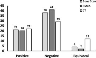
Region-based analysis revealed osseous metastatic regions in 78 (median 0 per patient, range 0–9), 25, and 87 (median 2 per patient, range 0–9) regions in the PSMA-11 scan, CT scan, and bone scan, respectively. CT scan was limited in assessing all nine osseous regions due to its restricted field of view.
The positive detection rate for local lymph-node and distant metastases (distant lymphatic, osseous, and visceral) was 18/63 (28.6%) and 23/63 (41.3%) for the PSMA-11 scan, and 20/63 (31.8%) and 27/63 (42.9%) for the CT scan, with no significant difference between the two modalities. Overall, the combined findings of the PSMA-11 scan, CT scan, and bone scan were positive in 31/63 (49.2%), 34/63 (53.9%), and 32/63 (50.8%) patients, respectively. Equivocal findings were reported in 1 PSMA-11 scan, 13 CT scans, and 4 bone scans. When equivocal findings were considered as positive for metastasis, the accuracy, sensitivity, and specificity were 78.2%, 60%, and 96.4% for the PSMA-11 scan; 76.1%, 62.9%, and 89.3% for the CT scan; and 85%, 78.6%, and 91.4% for the bone scan. There was a strong agreement in disease staging and overall findings between the PSMA-11 scan and the combination of CT and bone scans (Ƙ = 0.949 and Ƙ = 0.905, respectively; p < 0.001).
Conclusion
The comparable accuracy and high concordance between 99 mTc-HYNIC-PSMA-11 and conventional CT and bone scans make 99 mTc-HYNIC-PSMA-11 an effective method for initial staging of intermediate- to high-risk prostate cancer patients.
期刊介绍:
Annals of Nuclear Medicine is an official journal of the Japanese Society of Nuclear Medicine. It develops the appropriate application of radioactive substances and stable nuclides in the field of medicine.
The journal promotes the exchange of ideas and information and research in nuclear medicine and includes the medical application of radionuclides and related subjects. It presents original articles, short communications, reviews and letters to the editor.

 求助内容:
求助内容: 应助结果提醒方式:


