Paulina Truong, Saif Aldeen Alryalat, Osama Al Deyabat, Amina Malik, Masayoshi Takashima, Andrew Go Lee
{"title":"Orbital Fracture Resulting in Contralateral Optic Canal Fracture with Traumatic Optic Neuropathy: A Case Report.","authors":"Paulina Truong, Saif Aldeen Alryalat, Osama Al Deyabat, Amina Malik, Masayoshi Takashima, Andrew Go Lee","doi":"10.1159/000545040","DOIUrl":null,"url":null,"abstract":"<p><strong>Introduction: </strong>Traumatic optic neuropathy (TON) can cause acute vision loss after head trauma, either due to indirect shearing forces or direct trauma, i.e., by a bony fragment of an optic canal fracture (OCF). We present a case of TON due to an isolated OCF with contralateral orbital fracture and injury.</p><p><strong>Case presentation: </strong>A 19-year old male presented with immediate total loss of vision to no light perception in the right eye after being struck on the left cheek by a lawn sign. Computed tomography and magnetic resonance imaging revealed left orbital floor fracture and right optic nerve enhancement. The patient was treated with high-dose intravenous corticosteroids and plasma exchange for a presumed inflammatory or TON. Repeat orbital imaging revealed a right OCF with bony impingement of the optic nerve. The patient underwent endoscopic optic nerve decompression; a 4 × 5 mm bone fragment abutting the optic nerve was removed. 1 month later, vision improved to hand motion.</p><p><strong>Conclusion: </strong>Imaging may fail to detect OCF, and visual prognosis depends on time to surgery and fracture pattern. Therefore, operative management and preoperative intravenous corticosteroids, though controversial, may be considered even in the absence of radiographic findings of bony impingement causing direct TON. Isolated OCF without continuous fractures originating at the injury site is also a rare fracture pattern and potential cause of direct TON.</p>","PeriodicalId":9635,"journal":{"name":"Case Reports in Ophthalmology","volume":"16 1","pages":"290-296"},"PeriodicalIF":0.6000,"publicationDate":"2025-03-20","publicationTypes":"Journal Article","fieldsOfStudy":null,"isOpenAccess":false,"openAccessPdf":"https://www.ncbi.nlm.nih.gov/pmc/articles/PMC12021382/pdf/","citationCount":"0","resultStr":null,"platform":"Semanticscholar","paperid":null,"PeriodicalName":"Case Reports in Ophthalmology","FirstCategoryId":"1085","ListUrlMain":"https://doi.org/10.1159/000545040","RegionNum":0,"RegionCategory":null,"ArticlePicture":[],"TitleCN":null,"AbstractTextCN":null,"PMCID":null,"EPubDate":"2025/1/1 0:00:00","PubModel":"eCollection","JCR":"Q4","JCRName":"OPHTHALMOLOGY","Score":null,"Total":0}
引用次数: 0
Abstract
Introduction: Traumatic optic neuropathy (TON) can cause acute vision loss after head trauma, either due to indirect shearing forces or direct trauma, i.e., by a bony fragment of an optic canal fracture (OCF). We present a case of TON due to an isolated OCF with contralateral orbital fracture and injury.
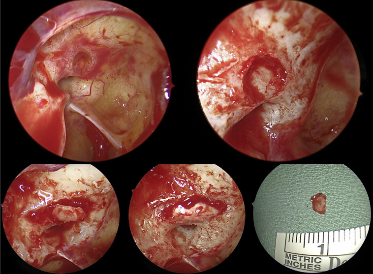
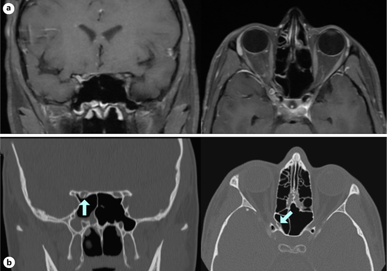
Case presentation: A 19-year old male presented with immediate total loss of vision to no light perception in the right eye after being struck on the left cheek by a lawn sign. Computed tomography and magnetic resonance imaging revealed left orbital floor fracture and right optic nerve enhancement. The patient was treated with high-dose intravenous corticosteroids and plasma exchange for a presumed inflammatory or TON. Repeat orbital imaging revealed a right OCF with bony impingement of the optic nerve. The patient underwent endoscopic optic nerve decompression; a 4 × 5 mm bone fragment abutting the optic nerve was removed. 1 month later, vision improved to hand motion.
Conclusion: Imaging may fail to detect OCF, and visual prognosis depends on time to surgery and fracture pattern. Therefore, operative management and preoperative intravenous corticosteroids, though controversial, may be considered even in the absence of radiographic findings of bony impingement causing direct TON. Isolated OCF without continuous fractures originating at the injury site is also a rare fracture pattern and potential cause of direct TON.
期刊介绍:
This peer-reviewed online-only journal publishes original case reports covering the entire spectrum of ophthalmology, including prevention, diagnosis, treatment, toxicities of therapy, supportive care, quality-of-life, and survivorship issues. The submission of negative results is strongly encouraged. The journal will also accept case reports dealing with the use of novel technologies, both in the arena of diagnosis and treatment. Supplementary material is welcomed. The intent of the journal is to provide clinicians and researchers with a tool to disseminate their personal experiences to a wider public as well as to review interesting cases encountered by colleagues all over the world. Universally used terms can be searched across the entire growing collection of case reports, further facilitating the retrieval of specific information. Following the open access principle, the entire contents can be retrieved at no charge, guaranteeing easy access to this valuable source of anecdotal information at all times.

 求助内容:
求助内容: 应助结果提醒方式:


