Alhasan H Alhebshi, Ammar Kabbarah, Murad Aljiffry
{"title":"Large Mesenchymal Hepatic Hamartoma in Pediatric Age: A Case Report.","authors":"Alhasan H Alhebshi, Ammar Kabbarah, Murad Aljiffry","doi":"10.1155/cris/1929050","DOIUrl":null,"url":null,"abstract":"<p><p>Benign liver tumors are infrequently observed in the pediatric age group, with an incidence reported at 0.7 per million population annually. Among these tumors, mesenchymal hamartoma constitutes 18%-29%. Imaging studies commonly reveal a well-marginated, solitary mass, often measuring up to 30 cm. The mass, primarily located in the right liver lobe (75% of cases), may exhibit a pedunculated structure. We present a case of a 1-year-and-9-month-old boy diagnosed with hepatic mesenchymal hamartoma. A contrast-enhanced computed tomography of the abdomen and magnetic resonance imaging (MRI) were performed and demonstrated a large multiloculated septated liver lesion measuring approximately 13.6 × 17.7 cm, demonstrating multiple partially thickened internal septations. The procedure was done for the patient in the form of an extended right hepatectomy with segment 4A and cholecystectomy.</p>","PeriodicalId":9600,"journal":{"name":"Case Reports in Surgery","volume":"2025 ","pages":"1929050"},"PeriodicalIF":0.5000,"publicationDate":"2025-04-30","publicationTypes":"Journal Article","fieldsOfStudy":null,"isOpenAccess":false,"openAccessPdf":"https://www.ncbi.nlm.nih.gov/pmc/articles/PMC12058315/pdf/","citationCount":"0","resultStr":null,"platform":"Semanticscholar","paperid":null,"PeriodicalName":"Case Reports in Surgery","FirstCategoryId":"1085","ListUrlMain":"https://doi.org/10.1155/cris/1929050","RegionNum":0,"RegionCategory":null,"ArticlePicture":[],"TitleCN":null,"AbstractTextCN":null,"PMCID":null,"EPubDate":"2025/1/1 0:00:00","PubModel":"eCollection","JCR":"Q4","JCRName":"SURGERY","Score":null,"Total":0}
引用次数: 0
Abstract
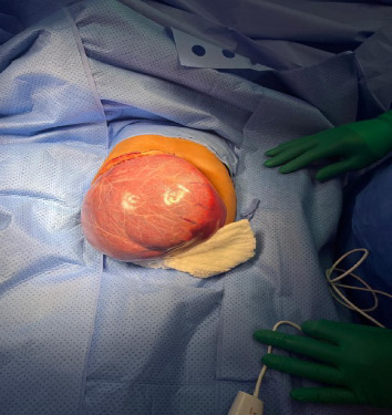
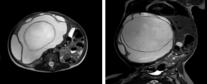
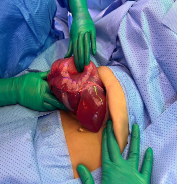
Benign liver tumors are infrequently observed in the pediatric age group, with an incidence reported at 0.7 per million population annually. Among these tumors, mesenchymal hamartoma constitutes 18%-29%. Imaging studies commonly reveal a well-marginated, solitary mass, often measuring up to 30 cm. The mass, primarily located in the right liver lobe (75% of cases), may exhibit a pedunculated structure. We present a case of a 1-year-and-9-month-old boy diagnosed with hepatic mesenchymal hamartoma. A contrast-enhanced computed tomography of the abdomen and magnetic resonance imaging (MRI) were performed and demonstrated a large multiloculated septated liver lesion measuring approximately 13.6 × 17.7 cm, demonstrating multiple partially thickened internal septations. The procedure was done for the patient in the form of an extended right hepatectomy with segment 4A and cholecystectomy.

 求助内容:
求助内容: 应助结果提醒方式:


