Gilbert M Schwarz, Alexander Synek, Stephanie Huber, Jochen G Hofstaetter, Dieter Pahr, Andreas Reisinger, Sylvia Nürnberger, Lena Hirtler
{"title":"Decreased femoral fracture load after cephalomedullary nail removal : a biomechanical ex vivo study.","authors":"Gilbert M Schwarz, Alexander Synek, Stephanie Huber, Jochen G Hofstaetter, Dieter Pahr, Andreas Reisinger, Sylvia Nürnberger, Lena Hirtler","doi":"10.1302/2046-3758.145.BJR-2024-0278.R2","DOIUrl":null,"url":null,"abstract":"<p><strong>Aims: </strong>Spontaneous neck fractures are feared complications of cephalomedullary nail removal after successful healing of per- and subtrochanteric fractures. To date, the initial postoperative stability as well as the correct weightbearing regimen remain unclear. The aim of this biomechanical ex vivo study was to evaluate the initial postoperative failure load after hardware removal of specimens, which received cephalomedullary nails during their lifetime.</p><p><strong>Methods: </strong>A total of 20 specimens of voluntary body donors were included in this study. Group 1 (n = 10) consisted of specimens that received cephalomedullary nails during their lifetime due to per- or subtrochanteric fractures. Each individual was matched for age, sex, femur size, and neck-shaft angle (Group 2 = control, n = 10). Biomechanical testing was performed in a single-leg stance setting, and volumetric bone mineral density (vBMD) was measured proximally at the femoral neck and distally at the epicondyles.</p><p><strong>Results: </strong>Groups 1 and 2 differed significantly in terms of failure loads (p = 0.002), fracture types, and ratios of proximal and distal vBMD (p = 0.035). Femora after nail removal were significantly weaker (1,835.0 N vs 4,523.0 N) and showed lower ratios of proximal to distal vBMD (0.74 vs 1.18), which indicated altered stress distributions at the femoral neck in presence of femoral neck screws. They were further characterized by predominantly subcapital buckle-type fractures, while the control Group 2 showed predominantly transcervical fractures.</p><p><strong>Conclusion: </strong>Altered stress distribution in presence of femoral neck screws leads to changes in biomechanical properties of the proximal femur, resulting in potentially unstable situations after nail removal in clinical settings. Elective removal of cephalomedullary nails should be undertaken with caution in view of the potentially increased fracture risk.</p>","PeriodicalId":9074,"journal":{"name":"Bone & Joint Research","volume":"14 5","pages":"368-375"},"PeriodicalIF":5.1000,"publicationDate":"2025-05-01","publicationTypes":"Journal Article","fieldsOfStudy":null,"isOpenAccess":false,"openAccessPdf":"https://www.ncbi.nlm.nih.gov/pmc/articles/PMC12043369/pdf/","citationCount":"0","resultStr":null,"platform":"Semanticscholar","paperid":null,"PeriodicalName":"Bone & Joint Research","FirstCategoryId":"3","ListUrlMain":"https://doi.org/10.1302/2046-3758.145.BJR-2024-0278.R2","RegionNum":2,"RegionCategory":"医学","ArticlePicture":[],"TitleCN":null,"AbstractTextCN":null,"PMCID":null,"EPubDate":"","PubModel":"","JCR":"Q2","JCRName":"CELL & TISSUE ENGINEERING","Score":null,"Total":0}
引用次数: 0
Abstract
Aims: Spontaneous neck fractures are feared complications of cephalomedullary nail removal after successful healing of per- and subtrochanteric fractures. To date, the initial postoperative stability as well as the correct weightbearing regimen remain unclear. The aim of this biomechanical ex vivo study was to evaluate the initial postoperative failure load after hardware removal of specimens, which received cephalomedullary nails during their lifetime.
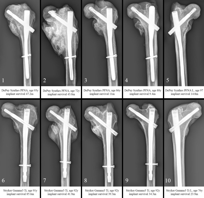
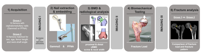
Methods: A total of 20 specimens of voluntary body donors were included in this study. Group 1 (n = 10) consisted of specimens that received cephalomedullary nails during their lifetime due to per- or subtrochanteric fractures. Each individual was matched for age, sex, femur size, and neck-shaft angle (Group 2 = control, n = 10). Biomechanical testing was performed in a single-leg stance setting, and volumetric bone mineral density (vBMD) was measured proximally at the femoral neck and distally at the epicondyles.
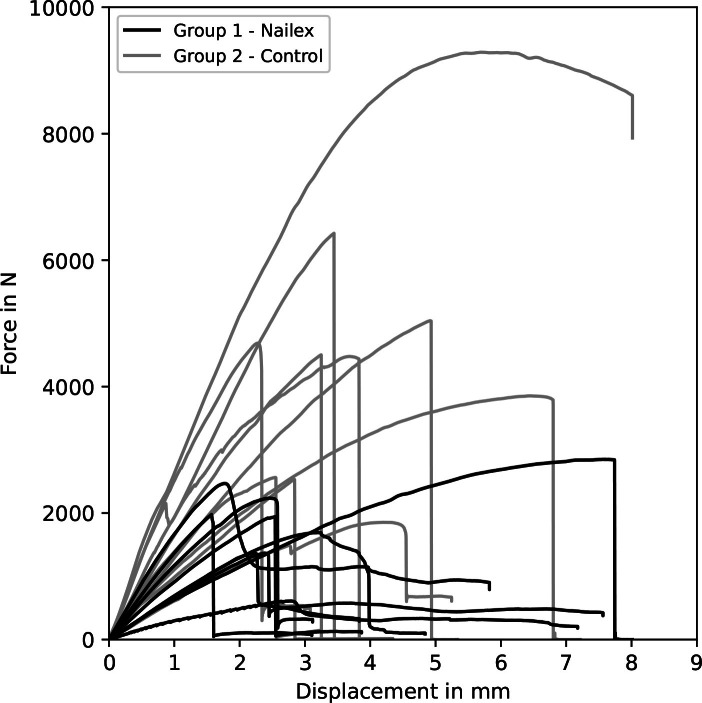
Results: Groups 1 and 2 differed significantly in terms of failure loads (p = 0.002), fracture types, and ratios of proximal and distal vBMD (p = 0.035). Femora after nail removal were significantly weaker (1,835.0 N vs 4,523.0 N) and showed lower ratios of proximal to distal vBMD (0.74 vs 1.18), which indicated altered stress distributions at the femoral neck in presence of femoral neck screws. They were further characterized by predominantly subcapital buckle-type fractures, while the control Group 2 showed predominantly transcervical fractures.
Conclusion: Altered stress distribution in presence of femoral neck screws leads to changes in biomechanical properties of the proximal femur, resulting in potentially unstable situations after nail removal in clinical settings. Elective removal of cephalomedullary nails should be undertaken with caution in view of the potentially increased fracture risk.
 求助内容:
求助内容: 应助结果提醒方式:


