{"title":"Sangyod Rice Extract Attenuates Vascular Inflammation and Injury in a Rat Model of Diabetes by Modulating the Akt/MAPK Signaling Pathway.","authors":"Wanwipha Woonnoi, Kornsuda Thipart, Wanthanee Hanchang, Jirawat Saetan, Supita Tanasawet, Furoida Moolsup, Wiwit Suttithumsatid, Tulaporn Wongtawatchai, Wanida Sukketsiri","doi":"10.1155/adpp/1169062","DOIUrl":null,"url":null,"abstract":"<p><p>Our previous study has shown the beneficial effect of the ethanolic extract of Sangyod rice (SE) on lipid accumulation and insulin resistance. However, its effect on vascular inflammation has yet to be explored. The current study aimed to investigate the anti-inflammatory effects of SE in both in vitro and in vivo models, specifically examining its impact on LPS-induced inflammation in RAW264.7 cells and evaluating its efficacy in an animal model of diabetes mellitus induced by a high-fat diet combined with a low-dose streptozotocin. In the in vitro experiments, SE treatment effectively suppressed the LPS-induced activation of key signaling pathways, including Akt, ERK1/2, p38 MAPK, and NF-κB, which are known to play pivotal roles in the inflammatory response. SE was also found to reduce oxidative stress and the production of inflammatory markers in the LPS-stimulated RAW264.7 cells. In the in vivo experiments, the administration of SE (500 mg/kg BW) and metformin (200 mg/kg BW) to high-fat diet/streptozotocin-induced diabetic rats effectively improved dyslipidemia, as evidenced by reductions in serum total cholesterol, LDL-cholesterol, and triglycerides compared to the untreated diabetic control group. Importantly, SE ameliorated the damage to the vascular endothelium and elastic fibers by downregulating the expression of proinflammatory cytokines and oxidative stress markers. Additionally, SE administration attenuated the upregulation of key markers associated with ER stress-mediated apoptotic pathways, with effects comparable to those observed in diabetic rats treated with the standard antidiabetic drug metformin. These findings suggest that SE possesses both anti-inflammatory and vascular protective properties, evident in both in vitro and in vivo studies.</p>","PeriodicalId":7369,"journal":{"name":"Advances in Pharmacological and Pharmaceutical Sciences","volume":"2025 ","pages":"1169062"},"PeriodicalIF":3.0000,"publicationDate":"2025-04-17","publicationTypes":"Journal Article","fieldsOfStudy":null,"isOpenAccess":false,"openAccessPdf":"https://www.ncbi.nlm.nih.gov/pmc/articles/PMC12021485/pdf/","citationCount":"0","resultStr":null,"platform":"Semanticscholar","paperid":null,"PeriodicalName":"Advances in Pharmacological and Pharmaceutical Sciences","FirstCategoryId":"1085","ListUrlMain":"https://doi.org/10.1155/adpp/1169062","RegionNum":0,"RegionCategory":null,"ArticlePicture":[],"TitleCN":null,"AbstractTextCN":null,"PMCID":null,"EPubDate":"2025/1/1 0:00:00","PubModel":"eCollection","JCR":"Q3","JCRName":"PHARMACOLOGY & PHARMACY","Score":null,"Total":0}
引用次数: 0
Abstract
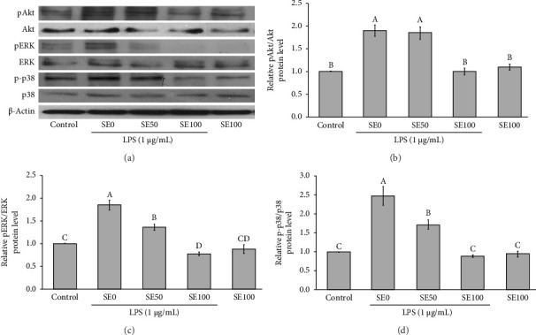
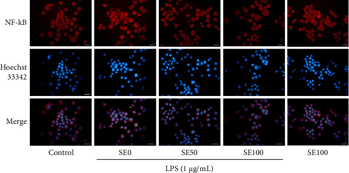
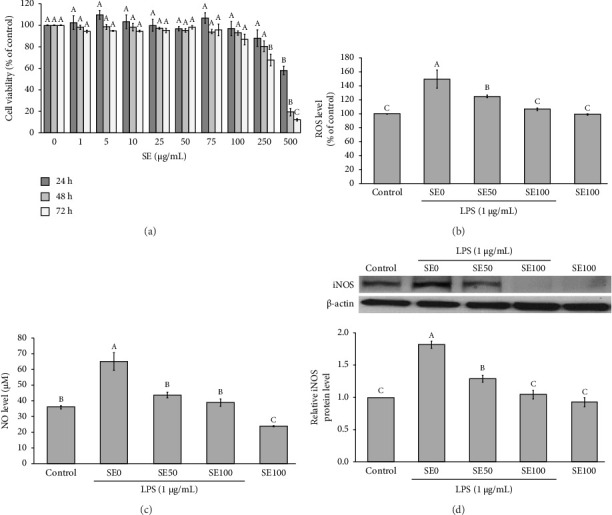
Our previous study has shown the beneficial effect of the ethanolic extract of Sangyod rice (SE) on lipid accumulation and insulin resistance. However, its effect on vascular inflammation has yet to be explored. The current study aimed to investigate the anti-inflammatory effects of SE in both in vitro and in vivo models, specifically examining its impact on LPS-induced inflammation in RAW264.7 cells and evaluating its efficacy in an animal model of diabetes mellitus induced by a high-fat diet combined with a low-dose streptozotocin. In the in vitro experiments, SE treatment effectively suppressed the LPS-induced activation of key signaling pathways, including Akt, ERK1/2, p38 MAPK, and NF-κB, which are known to play pivotal roles in the inflammatory response. SE was also found to reduce oxidative stress and the production of inflammatory markers in the LPS-stimulated RAW264.7 cells. In the in vivo experiments, the administration of SE (500 mg/kg BW) and metformin (200 mg/kg BW) to high-fat diet/streptozotocin-induced diabetic rats effectively improved dyslipidemia, as evidenced by reductions in serum total cholesterol, LDL-cholesterol, and triglycerides compared to the untreated diabetic control group. Importantly, SE ameliorated the damage to the vascular endothelium and elastic fibers by downregulating the expression of proinflammatory cytokines and oxidative stress markers. Additionally, SE administration attenuated the upregulation of key markers associated with ER stress-mediated apoptotic pathways, with effects comparable to those observed in diabetic rats treated with the standard antidiabetic drug metformin. These findings suggest that SE possesses both anti-inflammatory and vascular protective properties, evident in both in vitro and in vivo studies.

 求助内容:
求助内容: 应助结果提醒方式:


