Ilias Amtil-Ouahdi, Fabián Vergara, Carlos Rio, Coral González-Martínez, Andreas Jahn, María Antonia Forteza-Genestra, Antonio Gayá, Javier Calvo, Ernest Sala-Llinas, Bernardino Alcázar Navarrete, Ana Dolores Romero-Ortiz, Marta Monjo, Joana M. Ramis, Francisco G. Ortega
{"title":"EVs Biodistribution and Antifibrotic Impact in Aged Lung Fibrosis Model","authors":"Ilias Amtil-Ouahdi, Fabián Vergara, Carlos Rio, Coral González-Martínez, Andreas Jahn, María Antonia Forteza-Genestra, Antonio Gayá, Javier Calvo, Ernest Sala-Llinas, Bernardino Alcázar Navarrete, Ana Dolores Romero-Ortiz, Marta Monjo, Joana M. Ramis, Francisco G. Ortega","doi":"10.1002/biof.70021","DOIUrl":null,"url":null,"abstract":"<div>\n \n <p>Pulmonary fibrosis (PF) is a progressive, life-threatening disease marked by excessive scarring of lung tissue. Recently, extracellular vesicles (EVs) have emerged as a promising antifibrotic therapy due to their regenerative and anti-inflammatory properties. However, the success of EV-based therapies depends on the route of administration, which can significantly influence their biodistribution and therapeutic effects. Furthermore, in PF, aging is a significant risk factor for the disease and, until today, EV treatment efficacy has not been studied in aged tissues. Specifically, we studied EVs derived from human umbilical cord mesenchymal stem cells and compared the biodistribution of these vesicles delivered via three routes: intravenous (IV), intrapleural (IP), and intratracheal (IT). A protocol was developed to set EV staining and concentration, minimizing animal use while maximizing the accuracy of results. To evaluate therapeutic effects, we conducted three experimental setups: (i) to assess their ability to reverse established fibrosis; (ii) to evaluate their effect on fibrosis progression; and (iii) to study early inflammation and macrophage polarization. Lung fibrosis and inflammation were assessed by analyzing fibrotic markers, inflammatory cytokines, collagen deposition, and bronchoalveolar lavage (BAL) fluid cell analysis, providing insights into EVs therapeutic potential in aged, fibrotic lung tissue. In the biodistribution study, IV administration was identified as the most effective route, successfully delivering EVs to both normal and fibrotic lung tissues. In the therapeutic study, antifibrotic effects were observed only when EVs were administered prophylactically, before the establishment of fibrosis. Under this protocol, IV-administered EVs reduced fibrotic mRNA biomarkers, collagen deposition, inflammatory cell infiltration, and macrophage polarization in BAL, as well as altering cytokine. Our findings emphasize the critical importance of selecting the appropriate route of administration for EV-based therapies. Notably, our work with an aging model reveals that EV treatments primarily exhibit prophylactic effects, with a marked reduction in their regenerative potential compared to previous studies conducted in younger models.</p>\n </div>","PeriodicalId":8923,"journal":{"name":"BioFactors","volume":"51 3","pages":""},"PeriodicalIF":5.0000,"publicationDate":"2025-05-13","publicationTypes":"Journal Article","fieldsOfStudy":null,"isOpenAccess":false,"openAccessPdf":"","citationCount":"0","resultStr":null,"platform":"Semanticscholar","paperid":null,"PeriodicalName":"BioFactors","FirstCategoryId":"99","ListUrlMain":"https://iubmb.onlinelibrary.wiley.com/doi/10.1002/biof.70021","RegionNum":3,"RegionCategory":"生物学","ArticlePicture":[],"TitleCN":null,"AbstractTextCN":null,"PMCID":null,"EPubDate":"","PubModel":"","JCR":"Q1","JCRName":"BIOCHEMISTRY & MOLECULAR BIOLOGY","Score":null,"Total":0}
引用次数: 0
Abstract
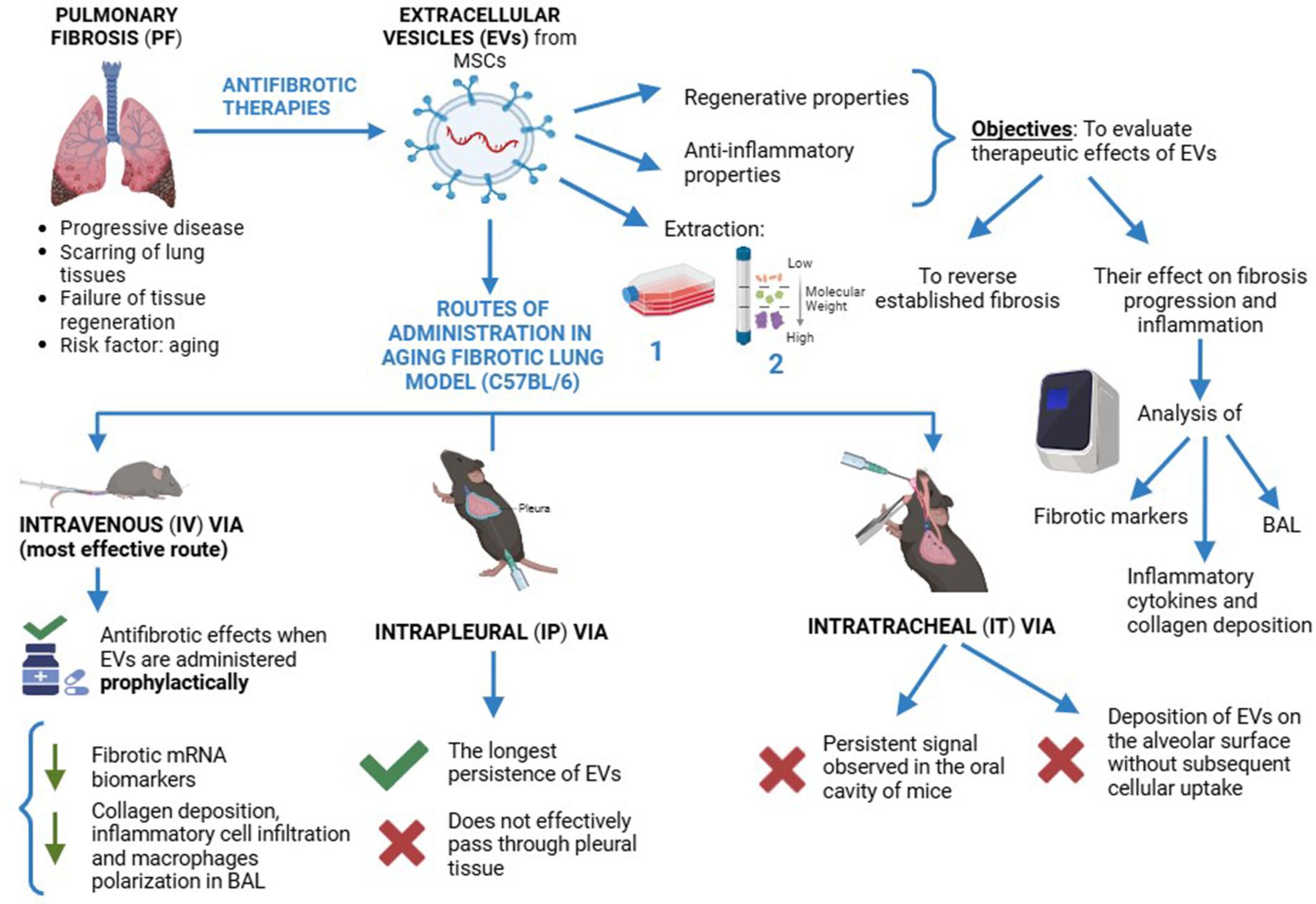
Pulmonary fibrosis (PF) is a progressive, life-threatening disease marked by excessive scarring of lung tissue. Recently, extracellular vesicles (EVs) have emerged as a promising antifibrotic therapy due to their regenerative and anti-inflammatory properties. However, the success of EV-based therapies depends on the route of administration, which can significantly influence their biodistribution and therapeutic effects. Furthermore, in PF, aging is a significant risk factor for the disease and, until today, EV treatment efficacy has not been studied in aged tissues. Specifically, we studied EVs derived from human umbilical cord mesenchymal stem cells and compared the biodistribution of these vesicles delivered via three routes: intravenous (IV), intrapleural (IP), and intratracheal (IT). A protocol was developed to set EV staining and concentration, minimizing animal use while maximizing the accuracy of results. To evaluate therapeutic effects, we conducted three experimental setups: (i) to assess their ability to reverse established fibrosis; (ii) to evaluate their effect on fibrosis progression; and (iii) to study early inflammation and macrophage polarization. Lung fibrosis and inflammation were assessed by analyzing fibrotic markers, inflammatory cytokines, collagen deposition, and bronchoalveolar lavage (BAL) fluid cell analysis, providing insights into EVs therapeutic potential in aged, fibrotic lung tissue. In the biodistribution study, IV administration was identified as the most effective route, successfully delivering EVs to both normal and fibrotic lung tissues. In the therapeutic study, antifibrotic effects were observed only when EVs were administered prophylactically, before the establishment of fibrosis. Under this protocol, IV-administered EVs reduced fibrotic mRNA biomarkers, collagen deposition, inflammatory cell infiltration, and macrophage polarization in BAL, as well as altering cytokine. Our findings emphasize the critical importance of selecting the appropriate route of administration for EV-based therapies. Notably, our work with an aging model reveals that EV treatments primarily exhibit prophylactic effects, with a marked reduction in their regenerative potential compared to previous studies conducted in younger models.
期刊介绍:
BioFactors, a journal of the International Union of Biochemistry and Molecular Biology, is devoted to the rapid publication of highly significant original research articles and reviews in experimental biology in health and disease.
The word “biofactors” refers to the many compounds that regulate biological functions. Biological factors comprise many molecules produced or modified by living organisms, and present in many essential systems like the blood, the nervous or immunological systems. A non-exhaustive list of biological factors includes neurotransmitters, cytokines, chemokines, hormones, coagulation factors, transcription factors, signaling molecules, receptor ligands and many more. In the group of biofactors we can accommodate several classical molecules not synthetized in the body such as vitamins, micronutrients or essential trace elements.
In keeping with this unified view of biochemistry, BioFactors publishes research dealing with the identification of new substances and the elucidation of their functions at the biophysical, biochemical, cellular and human level as well as studies revealing novel functions of already known biofactors. The journal encourages the submission of studies that use biochemistry, biophysics, cell and molecular biology and/or cell signaling approaches.


 求助内容:
求助内容: 应助结果提醒方式:


