The potential translational utility of embalmed cadaveric gastrointestinal tract specimens: a proof-of-concept study
Q3 Medicine
引用次数: 0
Abstract
Background
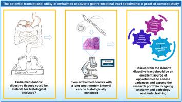
in recent decades, fewer autopsies are performed, often replaced by radiological imaging, resulting in abundant imaging data but few complete anatomical examinations. While most surgical specimens are limited to the disease-affected region. Examining the entire gastrointestinal system of anatomical donors and histologically analysing them could enhance research and valorise donation programs, extending medical knowledge. One factor limiting the preservation of the digestive tract in cadavers is the post-mortem interval. Embalming is one of the most widely used methods to preserve donors, but the post-mortem interval affects the gastrointestinal tract in a stable and time-dependent manner, making its histological examination a challenge.
Material and methods
this proof-of-concept study assesses the histological potential of gastrointestinal tissues from embalmed bodies used for anatomical education. Digestive tissues from four donors (two embalmed after 12 h, two after 72 h postmortem) were histologically processed. A scoring system evaluated histological preservation by assessing seven parameters.
Results
analysis showed high-quality preservation of embalmed tissues, including intestinal villi, the aorta, and liver sinusoids, highlighting both normal and pathological architecture, such as atherosclerosis, liver fibrosis, and lymphocyte infiltration. Features were identifiable with excellent histological detail in both the 12-h and 72-h post-mortem interval groups. Although this is a proof-of-concept study, only a slight difference was found between the two groups, with significant differences only in epithelium and vessel characteristics.
Conclusions
This study demonstrated that donors’ digestive tissues could be suitable for detailed analyses and insights into chronic diseases and ageing, even in cadavers with a long post-mortem interval.
尸体胃肠道标本防腐处理的潜在转化效用:一项概念验证研究
近几十年来,很少进行尸检,通常被放射成像所取代,导致大量的成像数据,但很少有完整的解剖检查。而大多数手术标本仅限于受疾病影响的区域。对解剖供体的整个胃肠道系统进行检查和组织学分析,可以加强研究,促进捐赠计划,扩展医学知识。限制尸体消化道保存的一个因素是死后的时间间隔。尸体防腐是最广泛使用的保存捐赠者的方法之一,但死后对胃肠道的影响是稳定的和时间依赖性的,这使得其组织学检查成为一个挑战。材料和方法本概念验证性研究评估了用于解剖学教育的防腐尸体胃肠道组织的组织学潜力。对4个供体的消化组织进行组织学处理(2个在死后12小时防腐处理,2个在死后72小时防腐处理)。评分系统通过评估七个参数来评估组织学保存。结果分析显示,高质量保存的防腐组织,包括肠绒毛、主动脉和肝窦,突出了正常和病理结构,如动脉粥样硬化、肝纤维化和淋巴细胞浸润。在死后12小时和72小时的时间间隔组中,这些特征都具有良好的组织学细节。虽然这是一项概念验证性研究,但两组之间仅发现轻微差异,仅在上皮和血管特征上存在显著差异。结论:这项研究表明,捐赠者的消化组织可以适用于对慢性疾病和衰老的详细分析和洞察,即使是在死后很长时间的尸体上。
本文章由计算机程序翻译,如有差异,请以英文原文为准。
求助全文
约1分钟内获得全文
求助全文
来源期刊

Translational Research in Anatomy
Medicine-Anatomy
CiteScore
2.90
自引率
0.00%
发文量
71
审稿时长
25 days
期刊介绍:
Translational Research in Anatomy is an international peer-reviewed and open access journal that publishes high-quality original papers. Focusing on translational research, the journal aims to disseminate the knowledge that is gained in the basic science of anatomy and to apply it to the diagnosis and treatment of human pathology in order to improve individual patient well-being. Topics published in Translational Research in Anatomy include anatomy in all of its aspects, especially those that have application to other scientific disciplines including the health sciences: • gross anatomy • neuroanatomy • histology • immunohistochemistry • comparative anatomy • embryology • molecular biology • microscopic anatomy • forensics • imaging/radiology • medical education Priority will be given to studies that clearly articulate their relevance to the broader aspects of anatomy and how they can impact patient care.Strengthening the ties between morphological research and medicine will foster collaboration between anatomists and physicians. Therefore, Translational Research in Anatomy will serve as a platform for communication and understanding between the disciplines of anatomy and medicine and will aid in the dissemination of anatomical research. The journal accepts the following article types: 1. Review articles 2. Original research papers 3. New state-of-the-art methods of research in the field of anatomy including imaging, dissection methods, medical devices and quantitation 4. Education papers (teaching technologies/methods in medical education in anatomy) 5. Commentaries 6. Letters to the Editor 7. Selected conference papers 8. Case Reports
 求助内容:
求助内容: 应助结果提醒方式:


