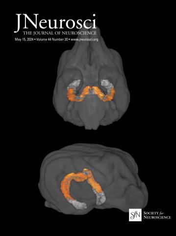
Differential impact of retinal lesions on visual responses of LGN X and Y cells.
IF 4.4
2区 医学
Q1 NEUROSCIENCES
引用次数: 0
Abstract
Damage to retinal cells from disease or injury causes vision loss and remodeling of downstream visual information processing circuits. As retinal cell replacement therapies and prosthetics become increasingly viable, we must understand post-retinal consequences of retinal cell loss to optimally recover visual perception. Here, we asked whether loss of retinal ganglion cells (RGCs) differentially impacts postsynaptic neurons in the visual thalamus - the dorsal lateral geniculate nucleus (LGN) - of ferrets, highly visual carnivores. We hypothesized that RGC loss might impact X more than Y LGN neurons, as there is less divergence in X retinogeniculate connections. We induced excitotoxic lesions of RGCs in a single eye and recorded neurophysiological responses of both contra- and ipsi-lesional LGN neurons to a variety of visual stimuli. We observed loss of responses among many LGN neurons, presumably with receptive fields within the scotoma. We also observed contralesional LGN neurons with receptive fields within or at the border of the scotoma that responded consistently to drifting sinusoidal gratings and spatiotemporally dynamic stimuli, enabling their classification as X or Y cells. Contralesional Y cell responses remained intact while contralesional X cells demonstrated higher firing rates, altered tuning to stimulus contrast and temporal frequency, and reduced spike timing precision. Consistent with neurophysiological results, alpha RGCs appeared relatively spared compared to beta RGCs. Together, our findings show that retinal cell loss differentially impacts downstream neuronal circuits, suggesting that supplemental vision recovery therapies may need to target visual circuits specialized for acuity vision.Significance Statement Vision loss from damage to retinal neurons may be partially ameliorated by improving cell replacement therapies and retinal prosthetics. However, retinal cell loss likely causes remodeling of circuits downstream, e.g., in the visual thalamus, which may also require treatment to restore natural visual perception. We studied neurophysiological changes in the visual thalamus, immediately postsynaptic to retina, following excitotoxic lesions of retinal output neurons. We discovered disproportionate effects on thalamic cell types, whereby cells receiving divergent retinal inputs were spared and those with few-to-one inputs had altered, noisier responses. Our findings suggest that supplemental vision therapies may need to target specific visual pathways to optimize acuity vision.视网膜病变对LGN X和Y细胞视觉反应的不同影响。
疾病或损伤对视网膜细胞的损害导致视力丧失和下游视觉信息处理回路的重塑。随着视网膜细胞替代疗法和假肢变得越来越可行,我们必须了解视网膜细胞损失的视网膜后后果,以最佳地恢复视觉感知。在这里,我们询问视网膜神经节细胞(RGCs)的损失是否会对视觉丘脑(背外侧膝状核(LGN))中的突触后神经元产生不同的影响,雪貂是高度视觉食肉动物。我们假设RGC丢失可能对X比Y LGN神经元的影响更大,因为X视网膜原化连接的分化较小。我们在一只眼睛中诱导RGCs的兴奋性毒性病变,并记录了对侧和同侧LGN神经元对各种视觉刺激的神经生理反应。我们观察到许多LGN神经元的反应丧失,可能与暗瘤内的接受野有关。我们还观察到,对侧LGN神经元在暗瘤内部或边界处具有接受野,它们对漂移正弦光栅和时空动态刺激做出一致的反应,从而使其分类为X或Y细胞。对侧Y细胞的反应保持不变,而对侧X细胞表现出更高的放电率,改变了对刺激对比度和时间频率的调节,降低了峰值定时精度。与神经生理学结果一致,与β RGCs相比,α RGCs显得相对幸免。总之,我们的研究结果表明,视网膜细胞损失对下游神经元回路的影响不同,这表明补充视力恢复疗法可能需要针对专门用于敏锐视力的视觉回路。通过改进细胞替代疗法和视网膜修复术,可以部分改善视网膜神经元损伤引起的视力丧失。然而,视网膜细胞的损失可能导致下游回路的重塑,例如视觉丘脑,这也可能需要治疗来恢复自然的视觉感知。我们研究了视网膜输出神经元兴奋性毒性损伤后,视觉丘脑突触后到视网膜的神经生理变化。我们发现了对丘脑细胞类型的不成比例的影响,即接受不同视网膜输入的细胞得以幸免,而那些接受少到一输入的细胞则产生了改变的、嘈杂的反应。我们的研究结果表明,补充视力疗法可能需要针对特定的视觉通路来优化锐度视力。
本文章由计算机程序翻译,如有差异,请以英文原文为准。
求助全文
约1分钟内获得全文
求助全文
来源期刊

Journal of Neuroscience
医学-神经科学
CiteScore
9.30
自引率
3.80%
发文量
1164
审稿时长
12 months
期刊介绍:
JNeurosci (ISSN 0270-6474) is an official journal of the Society for Neuroscience. It is published weekly by the Society, fifty weeks a year, one volume a year. JNeurosci publishes papers on a broad range of topics of general interest to those working on the nervous system. Authors now have an Open Choice option for their published articles
 求助内容:
求助内容: 应助结果提醒方式:


