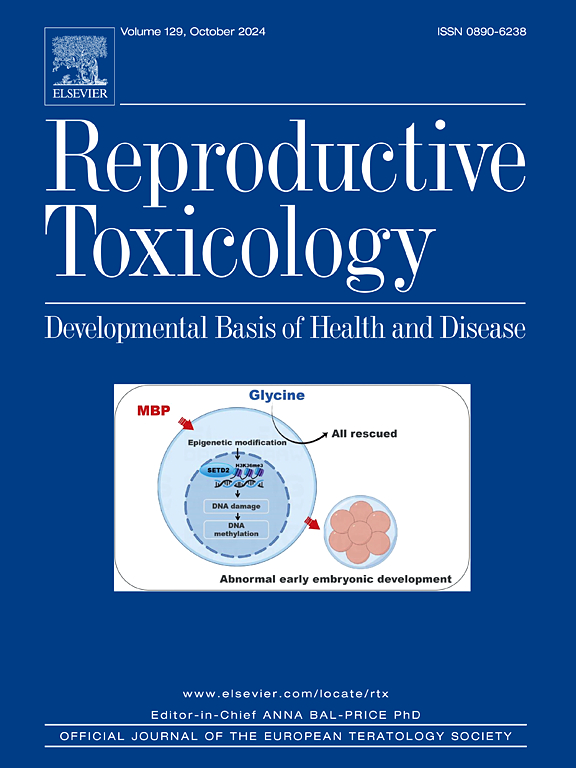
Maternal exposure to phthalates and nanoplastics, isolated or combined: Impacts on placental structure, development, and antioxidant defense as a trigger for maternal-fetal adversities
IF 2.8
4区 医学
Q2 REPRODUCTIVE BIOLOGY
引用次数: 0
Abstract
The placenta is an essential maternal-fetal organ for the healthy development of the fetus, linking maternal and fetal circulations. Endocrine disrupting chemicals (EDCs), such as phthalates derived from plastic residues, may impair offspring development and increase the risk of metabolic disorders. Plastics also degrade into microplastics (MPs) and nanoplastics (NPs), which can cross the placenta, carrying EDCs and impacting fetal development. The objective of this study was to investigate whether gestational exposure to a phthalate mixture (PM) and NPs interferes with the maternal-fetal interface, altering female reproductive efficiency and placental morphophysiology. Pregnant SD rats were divided into 6 groups: CTR(control; vehicle), T1(20 μg/kg/day-PM), T2(200 mg/kg/day-PM), T3(1 mg/kg/day NPs-100nm), T4(20 μg/kg/dayPM+1 mg/kg/day-NPs-100nm), and T5(200 mg/kg/day-PM+1 mg/kg/day-NPs-100 nm). Treatment was administered orally from gestational day 5 (GD5) to GD20. At GD20, 5–8 rats from each group were anesthetized and underwent laparotomy, and blood, ovaries, uterus, and placentas were analyzed. There was an increase in pre-implantation loss in T3, T4 and T5 groups, a reduction in placental weight, and an increase in placental efficiency in male offspring in T3 group. An increase in the number of fetuses small for gestational age was observed in T3 and T5 vs. C. Furthermore, the treatment caused an increase in the expression of targets related to trophoblast cell differentiation in T5, and growth factors related to angiogenesis in the placenta in T3 and T4 groups. There was a decrease in TBARS, SOD, and GSTpi levels in T2, while CAT increased in T3, suggesting that these pollutants modulate placental gene expression and energy metabolism.
母体接触邻苯二甲酸盐和纳米塑料,单独或联合:对胎盘结构、发育和抗氧化防御的影响,作为母胎逆境的触发因素
胎盘是胎儿健康发育必不可少的母胎器官,连接母体和胎儿的循环。内分泌干扰化学物质(EDCs),如来自塑料残留物的邻苯二甲酸盐,可能会损害后代的发育并增加代谢紊乱的风险。塑料也会降解成微塑料(MPs)和纳米塑料(NPs),它们可以穿过胎盘,携带EDCs并影响胎儿发育。本研究的目的是探讨妊娠期暴露于邻苯二甲酸盐混合物(PM)和NPs是否会干扰母胎界面,改变女性生殖效率和胎盘形态生理。将妊娠SD大鼠分为6组:CTR(对照组;T1(20 μg/kg/day- pm)、T2(200 mg/kg/day- pm)、T3(1 mg/kg/day NPs-100nm)、T4(20 mg/kg/day- pm +1 mg/kg/day-NPs-100nm)、T5(200 mg/kg/day- pm +1 mg/kg/day-NPs-100nm)。从妊娠第5天(GD5)至妊娠第20天口服治疗。GD20时,每组麻醉5-8只大鼠开腹,分析血液、卵巢、子宫、胎盘。T3、T4、T5组雄性子代胎盘着床前损失增加,胎盘重量减轻,胎盘效率提高。与c组相比,T3和T5组小胎数增加。此外,T3和T4组T5中与滋养细胞分化相关的靶细胞和胎盘中与血管生成相关的生长因子的表达增加。T2时TBARS、SOD和GSTpi水平下降,而T3时CAT水平升高,提示这些污染物调节了胎盘基因表达和能量代谢。
本文章由计算机程序翻译,如有差异,请以英文原文为准。
求助全文
约1分钟内获得全文
求助全文
来源期刊

Reproductive toxicology
生物-毒理学
CiteScore
6.50
自引率
3.00%
发文量
131
审稿时长
45 days
期刊介绍:
Drawing from a large number of disciplines, Reproductive Toxicology publishes timely, original research on the influence of chemical and physical agents on reproduction. Written by and for obstetricians, pediatricians, embryologists, teratologists, geneticists, toxicologists, andrologists, and others interested in detecting potential reproductive hazards, the journal is a forum for communication among researchers and practitioners. Articles focus on the application of in vitro, animal and clinical research to the practice of clinical medicine.
All aspects of reproduction are within the scope of Reproductive Toxicology, including the formation and maturation of male and female gametes, sexual function, the events surrounding the fusion of gametes and the development of the fertilized ovum, nourishment and transport of the conceptus within the genital tract, implantation, embryogenesis, intrauterine growth, placentation and placental function, parturition, lactation and neonatal survival. Adverse reproductive effects in males will be considered as significant as adverse effects occurring in females. To provide a balanced presentation of approaches, equal emphasis will be given to clinical and animal or in vitro work. Typical end points that will be studied by contributors include infertility, sexual dysfunction, spontaneous abortion, malformations, abnormal histogenesis, stillbirth, intrauterine growth retardation, prematurity, behavioral abnormalities, and perinatal mortality.
 求助内容:
求助内容: 应助结果提醒方式:


