Tissue and cell types infected by Ecytonucleospora hepatopenaei (EHP)

IF 2.4 3区 生物学 Q1 ZOOLOGY
Yuan Wang , Shi-ting Lin , Ming Zhao , Peng Di , Jun-fang Zhou , Shou-hu Li , Yan-qing Huang , Ying Na , Xin-cang Li , Wen-hong Fang
{"title":"Tissue and cell types infected by Ecytonucleospora hepatopenaei (EHP)","authors":"Yuan Wang ,&nbsp;Shi-ting Lin ,&nbsp;Ming Zhao ,&nbsp;Peng Di ,&nbsp;Jun-fang Zhou ,&nbsp;Shou-hu Li ,&nbsp;Yan-qing Huang ,&nbsp;Ying Na ,&nbsp;Xin-cang Li ,&nbsp;Wen-hong Fang","doi":"10.1016/j.jip.2025.108344","DOIUrl":null,"url":null,"abstract":"<div><div><em>Ecytonucleospora hepatopenaei</em> (EHP) is a microsporidian pathogen causing significant losses in shrimp aquaculture worldwide. The hepatopancreas is recognized as the primary target tissue, but the broader tissue and cell tropism of EHP and its ability to infect other components of the digestive system or non-digestive tissues remain unclear, especially when infections are intense and the host physiology is compromised. This study aimed to comprehensively investigate the histopathology of EHP infections in severely infected <em>Penaeus vannamei</em> to determine its tissue and cell tropism and assess the potential for systemic infection. The severity of infection was graded based on hepatopancreatic lesions. Histopathology showed that EHP spores were distributed in the digestive system of heavily infected shrimp, but not in non-digestive tissues such as gills, heart, gonad, nerves or skeletal muscle. EHP only infected the epithelial cells of the hepatopancreas, midgut, and midgut caeca, which lack the protective chitin layers. While the epithelial cells of the esophagus, stomach and hindgut were unaffected due to the protection of the inner chitinous layer, despite the presence of large numbers of EHP spores in these regions. Histopathology and ultrastructural pathology demonstrated that the R (reserve), F (fibrillar), B (blister), E (embryonic) and M (small midget) cells of the hepatopancreas were infected. These findings indicate that EHP does not cause systemic infection and has a strict cell tropism for the epithelium in the shrimp host.</div></div>","PeriodicalId":16296,"journal":{"name":"Journal of invertebrate pathology","volume":"211 ","pages":"Article 108344"},"PeriodicalIF":2.4000,"publicationDate":"2025-04-26","publicationTypes":"Journal Article","fieldsOfStudy":null,"isOpenAccess":false,"openAccessPdf":"","citationCount":"0","resultStr":null,"platform":"Semanticscholar","paperid":null,"PeriodicalName":"Journal of invertebrate pathology","FirstCategoryId":"99","ListUrlMain":"https://www.sciencedirect.com/science/article/pii/S0022201125000783","RegionNum":3,"RegionCategory":"生物学","ArticlePicture":[],"TitleCN":null,"AbstractTextCN":null,"PMCID":null,"EPubDate":"","PubModel":"","JCR":"Q1","JCRName":"ZOOLOGY","Score":null,"Total":0}
引用次数: 0

Abstract

Ecytonucleospora hepatopenaei (EHP) is a microsporidian pathogen causing significant losses in shrimp aquaculture worldwide. The hepatopancreas is recognized as the primary target tissue, but the broader tissue and cell tropism of EHP and its ability to infect other components of the digestive system or non-digestive tissues remain unclear, especially when infections are intense and the host physiology is compromised. This study aimed to comprehensively investigate the histopathology of EHP infections in severely infected Penaeus vannamei to determine its tissue and cell tropism and assess the potential for systemic infection. The severity of infection was graded based on hepatopancreatic lesions. Histopathology showed that EHP spores were distributed in the digestive system of heavily infected shrimp, but not in non-digestive tissues such as gills, heart, gonad, nerves or skeletal muscle. EHP only infected the epithelial cells of the hepatopancreas, midgut, and midgut caeca, which lack the protective chitin layers. While the epithelial cells of the esophagus, stomach and hindgut were unaffected due to the protection of the inner chitinous layer, despite the presence of large numbers of EHP spores in these regions. Histopathology and ultrastructural pathology demonstrated that the R (reserve), F (fibrillar), B (blister), E (embryonic) and M (small midget) cells of the hepatopancreas were infected. These findings indicate that EHP does not cause systemic infection and has a strict cell tropism for the epithelium in the shrimp host.

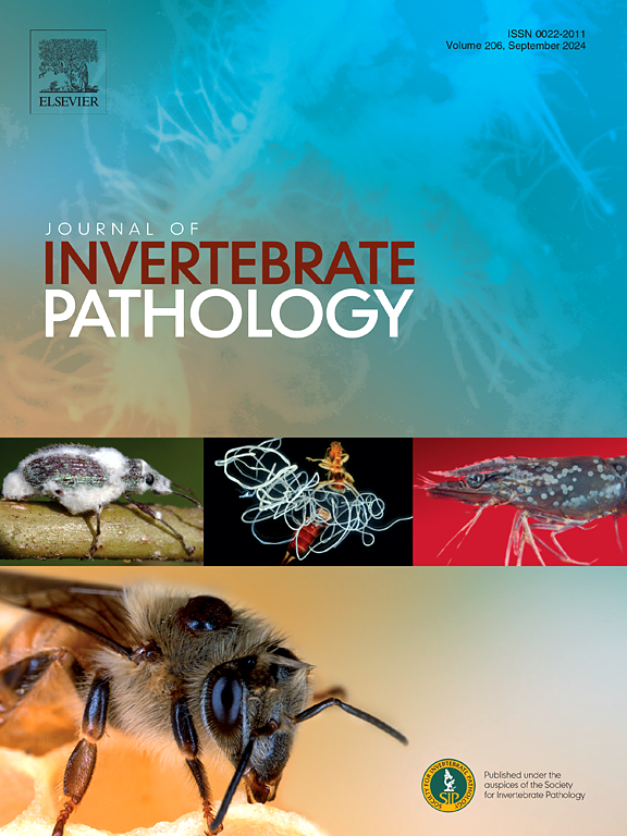
Abstract Image

肝外核孢子虫(EHP)感染组织和细胞类型
肝原胞核孢子虫(EHP)是一种微孢子虫致病菌,在全球对虾养殖中造成重大损失。肝胰腺被认为是主要的目标组织,但EHP更广泛的组织和细胞趋向性及其感染消化系统其他成分或非消化组织的能力尚不清楚,特别是当感染强烈且宿主生理受损时。本研究旨在全面研究严重感染的凡纳滨对虾EHP感染的组织病理学,以确定其组织和细胞的嗜性,并评估其全身性感染的可能性。感染的严重程度根据肝胰脏病变程度分级。组织病理学结果显示,重感染对虾的消化系统中存在EHP孢子,而鳃、心脏、性腺、神经和骨骼肌等非消化组织中不存在孢子。EHP仅感染肝胰腺、中肠和中肠盲肠的上皮细胞,这些细胞缺乏几丁质保护层。而食管、胃和后肠的上皮细胞则不受影响,尽管这些区域存在大量的EHP孢子,但由于内层几质层的保护。组织病理学和超微结构病理学显示肝胰腺R(储备细胞)、F(纤维细胞)、B(水疱细胞)、E(胚胎细胞)和M(小细胞)受到感染。这些结果表明EHP不会引起全身感染,并且对宿主虾的上皮具有严格的细胞亲和性。
本文章由计算机程序翻译,如有差异,请以英文原文为准。
求助全文
约1分钟内获得全文 求助全文
来源期刊
CiteScore
6.10
自引率
5.90%
发文量
94
审稿时长
1 months
期刊介绍: The Journal of Invertebrate Pathology presents original research articles and notes on the induction and pathogenesis of diseases of invertebrates, including the suppression of diseases in beneficial species, and the use of diseases in controlling undesirable species. In addition, the journal publishes the results of physiological, morphological, genetic, immunological and ecological studies as related to the etiologic agents of diseases of invertebrates. The Journal of Invertebrate Pathology is the adopted journal of the Society for Invertebrate Pathology, and is available to SIP members at a special reduced price.
×
引用
GB/T 7714-2015
复制
MLA
复制
APA
复制
导出至
BibTeX EndNote RefMan NoteFirst NoteExpress
×
提示
您的信息不完整,为了账户安全,请先补充。
现在去补充
×
提示
您因"违规操作"
具体请查看互助需知
我知道了
×
提示
确定
请完成安全验证×
copy
已复制链接
快去分享给好友吧!
我知道了
右上角分享
点击右上角分享
0
联系我们:info@booksci.cn Book学术提供免费学术资源搜索服务,方便国内外学者检索中英文文献。致力于提供最便捷和优质的服务体验。 Copyright © 2023 布克学术 All rights reserved.
京ICP备2023020795号-1
ghs 京公网安备 11010802042870号
Book学术文献互助
Book学术文献互助群
群 号:604180095
Book学术官方微信